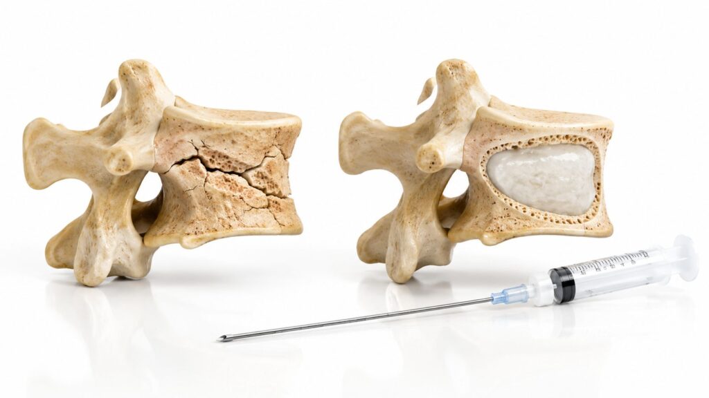
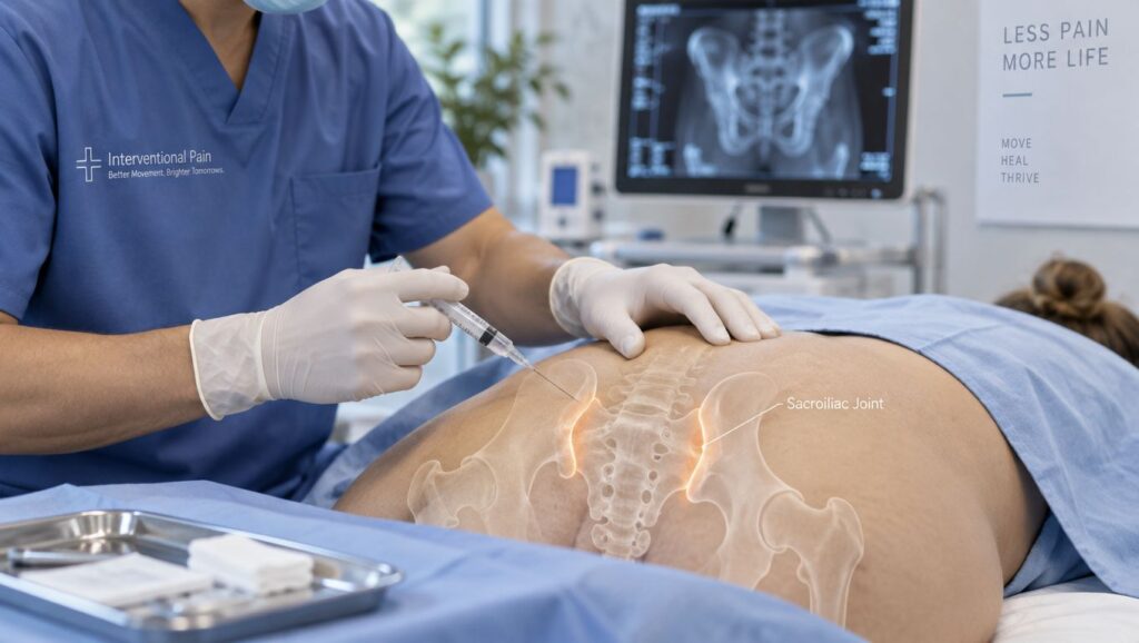
You've tried to solve your “back pain” from every angle. You've had imaging, perhaps an epidural injection, and a course of physical therapy, yet a very specific ache near one side of your pelvis still wakes you when you roll in bed. That pattern can be frustrating, especially when the pain doesn't behave like the disc or nerve problem suggested by earlier treatments.
Radiofrequency ablation for the SI joint may help some carefully selected patients, but it isn't a guaranteed fix and it isn't appropriate for every type of sacroiliac pain. The key questions are whether the SI joint is generating your symptoms, whether a diagnostic block supports that conclusion, which RFA technique is being considered, and whether your insurance policy allows access to it.
Table of Contents
- When the Pain in Your Lower Back Isn't Really Your Back
- The SI Joint and Why It Causes So Much Trouble
- Who Is a Real Candidate for SI Joint RFA
- What Happens During the Procedure
- What the Evidence Shows
- Coverage Rules and the Access Gap
- How RFA Fits Into a Multimodal Pain Plan
When the Pain in Your Lower Back Isn't Really Your Back
A desk worker points to one spot beside the middle of her lower back. Rolling in bed, climbing stairs, or standing mainly on one leg makes it worse. She has already undergone two MRI scans, tried an epidural steroid injection, and completed physical therapy, yet the same area continues to hurt.
That pattern can involve the sacroiliac joint, commonly called the SI joint. It sits where the lower spine meets the pelvis, so pain from this region can look like lumbar disc disease, facet pain, or nerve-root irritation. The discomfort may be felt in the buttock, hip, or low back, even when the lumbar structures targeted by an epidural are not the main pain source.
Broader reviews estimate that SI joint pain accounts for up to 25% of low back pain cases, rising to 32% to 37% after lumbosacral fusion surgery (The clinical evidence on SI joint RFA and its patient-selection context). These figures help explain why a patient may continue receiving treatments aimed at the lumbar spine while the pain generator is closer to the pelvis. They do not prove that the SI joint is responsible in any one person. Diagnosis still depends on the history, examination, and response to targeted testing.

Why the location matters
The SI joint moves only slightly, but it transfers force between the spine and legs during walking, standing, twisting, and climbing. Pain can stay near the joint or travel into the buttock and upper leg. That spread makes self-diagnosis unreliable, especially for someone who has already had lumbar imaging or treatment.
In an Illinois pain clinic, a physician typically combines the pain pattern, physical examination, and response to a controlled diagnostic injection. If numbing the suspected joint region temporarily removes the familiar pain, radiofrequency ablation SI joint treatment may become a targeted option to discuss. RFA interrupts selected pain signals. It does not repair the joint or address every possible cause of lower-back pain, so it belongs within a broader, individualized plan rather than serving as a guaranteed fix.
The SI Joint and Why It Causes So Much Trouble
Think of your pelvis as a solid foundation and your spine as a loaded barge tied to it. The two SI joints act like small docks between them. They aren't designed for large movements, but they absorb forces from walking, lifting, turning, and transferring weight from one leg to the other.
That arrangement can become painful after an injury, pregnancy-related changes, lumbar fusion, inflammation, or long-term uneven loading. A stiff hip, weak pelvic stabilizers, or altered walking pattern can also increase stress around the joint. The important point is that the painful structure may be the joint complex and its surrounding ligaments, not a lumbar nerve root.
What RFA actually targets
The posterior SI joint region receives sensory input from the L5 dorsal ramus and the S1 through S3 lateral branches. These nerves carry pain signals from the structures around the joint. RFA applies controlled energy near selected nerve targets to reduce their ability to transmit those signals.
That's different from an epidural injection, which is intended for inflammation around spinal nerve roots. A disc-directed treatment may be useful when a disc or lumbar nerve is the pain generator, but it won't directly treat pain arising from the SI joint region.
Before considering ablation, it helps to understand the broader range of ways clinicians may treat SI joint pain. Options can include activity changes, targeted rehabilitation, injections, and procedures aimed at the sensory nerves. For a description of local evaluation and treatment pathways, review these sacroiliac joint pain treatment options.
The practical distinction
RFA doesn't fuse the joint, realign the pelvis, or remove a disc. It's a pain-modulating procedure. That makes accurate diagnosis essential, because reducing sensation from the wrong nerve targets won't correct the actual problem.
Practical rule: The more closely your temporary diagnostic relief matches your usual pain, the more useful that block is for deciding whether nerve-targeted treatment makes sense.
Who Is a Real Candidate for SI Joint RFA
Candidacy should develop in stages rather than being decided from an MRI alone. Imaging may show arthritis, degeneration, or postoperative changes, but those findings don't prove that the SI joint is causing your current symptoms.
Start with the clinical pattern
A clinician looks for pain concentrated near the back of the pelvic brim, along with symptoms reproduced by specific maneuvers. Common examination tools include the Patrick's test and thigh thrust test, along with other SI-joint provocation maneuvers. The goal isn't to pass one isolated test. It's to see whether several findings point toward the same pain generator.
Conservative treatment generally comes first. That may include pelvic-focused physical therapy, activity modification, medication when medically appropriate, and an image-guided SI joint injection that provides both diagnostic information and a therapeutic trial.
The diagnostic block is the gatekeeper
RFA targets sensory nerves rather than the joint space itself, so clinicians may use fluoroscopic blocks of the L5 dorsal ramus and S1 through S3 lateral branches. During a block, a small amount of local anesthetic temporarily interrupts the targeted signals. You'll be asked to compare your familiar pain during the anesthetic window with your usual symptoms during the same activities.
The most persuasive result is relief that is substantial, temporary, and functionally meaningful. A patient who can roll in bed, stand, or walk with much less of the familiar pain provides more useful information than someone who reports only vague improvement.
Coverage policies don't use one universal standard. Some policies require 75% or greater relief after a diagnostic injection, while a 2025 multidisciplinary consensus discussed sacral lateral branch blocks with a 50% cutoff as a prognostic approach before lateral branch RFA. The current payer-policy document and diagnostic-threshold discussion illustrate why a clinician's recommendation and an insurer's approval decision may differ.
When RFA may not be appropriate
Active infection, an untreated bleeding disorder, inability to safely stop certain blood-thinning medicines, or uncontrolled psychiatric illness can make an elective procedure unsafe or unsuitable. A clinician should also reconsider RFA when symptoms are better explained by a hip disorder, lumbar nerve compression, inflammatory disease, fracture, or another condition requiring different treatment.
What Happens During the Procedure
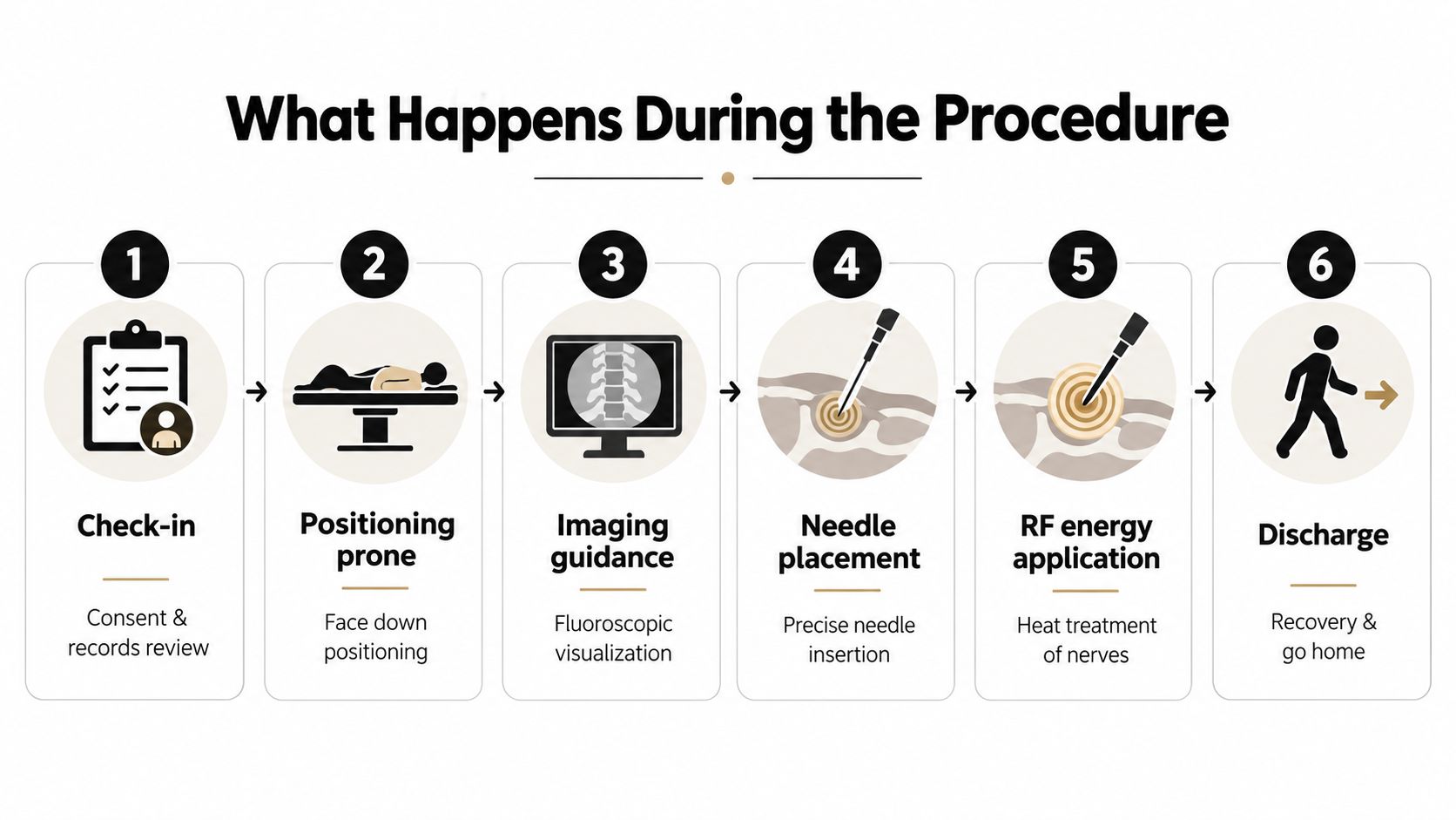
You'll usually begin with consent, a review of your medical history, and confirmation of the diagnostic-block results. Tell the team about blood thinners, infection symptoms, medication allergies, and any change in your pain pattern since the block.
You'll lie face down on the procedure table. The clinician cleans the skin over the back of the pelvis and uses local anesthetic to numb the entry points. Fluoroscopy provides live X-ray guidance so the treatment needles can be directed toward the L5 dorsal ramus and the S1 through S3 lateral branch targets.

What you may feel
After the needle reaches the intended position, the clinician may perform brief sensory and motor testing. You might notice tingling, tapping, or pressure. This testing helps confirm that the electrode is near the intended sensory target and away from an important motor nerve.
For conventional thermal RFA, the electrode creates a controlled heat lesion. The treatment plan may use heat around 80°C for roughly 60 to 90 seconds per site, but the exact settings depend on the technique, equipment, anatomy, and physician judgment. Those technical details should be explained during consent rather than treated as a promise of a specific result.
The procedure can feel like pressure or warmth more than sharp pain. If you feel significant discomfort, tell the clinician. Local anesthetic is used to make the treatment more tolerable, and some patients receive medication to reduce anxiety when medically appropriate.
Leaving the clinic
Afterward, staff monitor you during recovery. A 30-to-60-minute observation period may be typical, though the actual time depends on sedation, medical history, and how you feel. You'll generally need a responsible adult to help you get home if sedating medication was used.
The treated area can be sore before the nerve-calming effect becomes clear. Temporary numbness, tenderness, or a short-lived increase in pain can occur. Follow the discharge instructions, avoid judging the result on the same day, and record changes in pain and function over the following days.
For more on why imaging matters during injections and other procedures, see this explanation of fluoroscopy-guided injection.
What the Evidence Shows
The evidence supports cautious optimism, not a guarantee. SI joint RFA can reduce pain for selected patients, but results depend on confirming the pain source, placing the treatment accurately, and choosing a technique that matches the nerve pattern being treated. The procedure is better understood as turning down one pain signal, not repairing every possible cause of pain around the pelvis.
A foundational randomized placebo-controlled study included 28 patients. 14 received lateral branch radiofrequency denervation, and 14 received placebo denervation. In the active-treatment group, at least 50% pain relief and functional improvement occurred in 79% at one month, 64% at three months, and 57% at six months. The historical trial and earlier technique reports helped establish that improvement could extend beyond a sham procedure, while also showing that treatment methods have changed over time.
What newer comparative evidence adds
A multicenter randomized study of 210 patients compared cooled RFA with standard medical management. At three months, the mean pain score was 3.8±2.4 with cooled RFA versus 5.9±1.7 with standard care. Pain reduction was 2.5±2.5 versus 0.4±1.7, and the responder rate was 52.3% versus 4.3%, using a definition of more than two points or 30% pain relief. The comparative-effectiveness study offers a useful benchmark, but it does not predict the result for every individual.
At 12 months in the same randomized program, the mean NRS decreased from 6.4±1.4 to 3.5±2.6, and 57.4% met the threshold of at least two points or 30% improvement after a single treatment. A 2024 systematic review described the overall evidence as Level III with a fair recommendation, found no major adverse events in its pooled comparison, and found no statistical efficacy difference between conventional and cooled RFA across nine studies and 276 patients. The longer-term review and meta-analysis also emphasizes that the evidence remains limited.
SI Joint RFA techniques compared
| Technique | Lesion Size | Reported Success Rate | Average Duration of Relief |
|---|---|---|---|
| Conventional or monopolar RFA | Varies by electrode and settings. No single size is established in the supplied evidence. | Results vary by technique and study, so one dependable rate cannot be assigned. | Relief can be temporary, and durability varies by study. |
| Bipolar RFA | Early reports used bipolar electrodes. A standardized lesion size is not established in the supplied evidence. | Sustained six-month relief was reported in 36% in an early technique series. Early bipolar technique series (Cohen et al.) | Six-month relief was reported in that early series. |
| Cooled RFA | Designed to create a larger treatment zone than some conventional approaches, but no single lesion size should be assumed. | 52.3% responded at three months, and 57.4% met the improvement threshold at 12 months in the same randomized program. Randomized comparative program, 3- and 12-month outcomes. | Benefit was documented through 12 months in selected patients. |
| Pulsed RFA | No definitive lesion-size figure is established in the supplied evidence. | A dependable pooled rate is not established in the supplied evidence. | A dependable average is not established in the supplied evidence. |
A 2026 systematic review found that conventional lateral branch RFA did not show a specific treatment effect in some sham-controlled SI-joint trials, while strip-lesion, capsular, or cooled approaches produced greater and more sustained relief in selected studies. A separate network meta-analysis of 18 trials and 1,075 patients found RF-based interventions outperformed steroid injections, sham, and conservative care overall, but rated certainty as low to very low because of imprecision and study limitations. The 2026 review and network analysis explain why technique and patient selection matter as much as the label “RFA.” The practical question is whether the pain comes from the treated SI joint complex rather than another generator.
Coverage Rules and the Access Gap
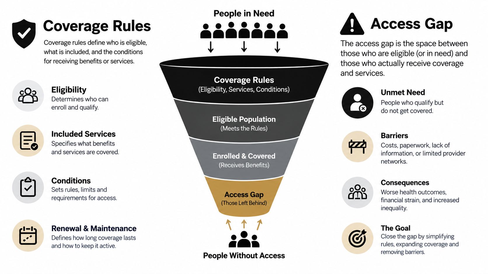
A physician may judge SI joint RFA clinically reasonable while an insurer still refuses authorization. Approval depends on documentation, not only on the treatment decision. Payers commonly request the pain pattern, examination findings, imaging review, prior conservative care, response to injections, and diagnostic-block results, often in a specified sequence.
Some policies classify SI joint RFA as experimental, investigational, or unproven. Others require a documented high response threshold after a diagnostic injection and limit how often repeat treatment may occur. These rules can differ from the threshold a clinician uses when deciding whether the SI joint is contributing to a patient's pain.
Why one block can create confusion
A diagnostic block is a short test, not a permanent treatment. Its result can be affected by whether the medication reached the intended target, which activity the patient used for comparison, and whether another pain generator was active that day. A patient may therefore report meaningful but incomplete relief, even when the SI joint is part of the pain pattern.
A 2025 consensus discussion considered sacral lateral branch blocks with a lower cutoff before lateral branch RFA, while payer policies may demand a higher response. That difference explains why a clinician may recommend proceeding after meaningful temporary relief while an insurer requests another block or denies authorization. The disagreement often concerns the coverage rule, not whether the patient's symptoms are real.
How the process usually unfolds
The clinic submits clinical notes, procedure reports, medication history, examination findings, and block results for prior authorization. The insurer may request additional records or a peer-to-peer review. If authorization is denied, the care team can examine the stated reason, correct missing documentation, and submit an appeal when appropriate.
Request the coverage criteria in writing before scheduling RFA. Reviewing radiofrequency ablation cost can also separate insurance approval from facility charges, professional fees, and possible out-of-pocket responsibility. These are different questions, and approval of the procedure does not always clarify the final patient balance.
Local access can also affect which clinic a patient chooses. Patients in Chicago Ridge, Palos Heights, Palos Hills, Evergreen Park, and Hickory Hills are within a short drive of one another, making it practical to compare local care options. The local geographic reference provides context for these nearby Illinois communities.
How RFA Fits Into a Multimodal Pain Plan
RFA works best as one layer in a broader plan. It can reduce pain transmission from selected nerves, but it won't automatically correct weak pelvic stabilization, poor movement mechanics, hip dysfunction, inflammatory disease, or a separate lumbar problem.
A practical pathway often begins with targeted physical therapy, activity modification, and medication when appropriate. Therapy may focus on pelvic control, hip strength, trunk coordination, and movements that let you load one leg without repeatedly provoking the joint.
Choosing the next layer
An image-guided SI joint injection may serve as a therapeutic trial and help clarify the pain source. If relief is temporary but clearly matches the familiar pain, the clinician may discuss RFA. A 2022 systematic review concluded that the available evidence supports SI joint RFA for chronic pain for periods of up to one year after treatment, which gives a useful published boundary without promising that every patient will reach it. The systematic review on SI joint RFA supports setting expectations around temporary, potentially repeatable relief.
Platelet-rich plasma and other regenerative approaches may be discussed in selected cases, but their durability remains less certain. Neuromodulation, including peripheral nerve stimulation or spinal cord stimulation, may enter the conversation when pain persists despite other interventions. SI joint fusion is a separate surgical decision, generally reserved for carefully confirmed pain that remains disabling despite nonsurgical care.
Sleep deserves attention during every stage. Pain can make turning in bed difficult, and poor sleep can make pain harder to manage. Patients looking for practical strategies can review this guide to find sleep relief from chronic pain.
A reasonable Illinois care plan
Bring your imaging, procedure reports, medication list, and a short record of activities that trigger or relieve pain. Then ask:
- Source confirmation: Which findings suggest the SI joint, rather than the lumbar spine or hip, is responsible?
- Diagnostic testing: Which block is being recommended, and what result would support moving forward?
- Technique choice: Why is conventional, cooled, bipolar, or another approach being considered?
- Coverage preparation: What documentation does your Illinois insurance policy require?
- Functional goal: Will success mean sleeping better, walking farther, returning to work, or reducing medication?
A Chicago Ridge pain clinic can coordinate evaluation, diagnostic injections, rehabilitation referrals, and image-guided procedures as parts of one plan. The purpose is not just to reduce a pain score. It's to help you move, sleep, work, and participate in daily life with less reliance on escalating medication.
Midwest Pain & Wellness offers evaluation and multimodal treatment for SI joint and other chronic pain conditions, including diagnostic injections and radiofrequency ablation when clinically appropriate. Visit Midwest Pain & Wellness to request a consultation serving Chicago Ridge, Palos Heights, Palos Hills, Evergreen Park, Hickory Hills, and nearby Illinois communities.