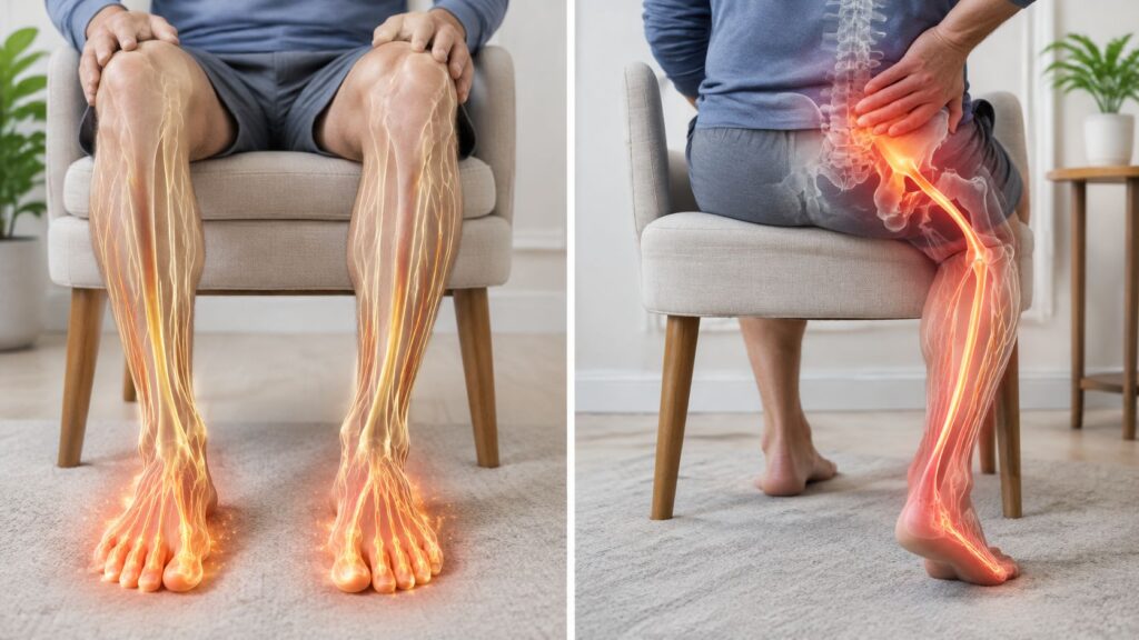
You've had low back or buttock pain for months. You've tried medication, exercises, and perhaps physical therapy. A lumbar MRI may not have explained why sitting, standing, climbing stairs, or turning in bed still hurts. At that point, it's reasonable to ask, what is a sacroiliac joint injection, and could it both identify the painful structure and calm it down?
An SI joint injection is an image-guided procedure that places medication into the sacroiliac joint, where the lower spine meets the pelvis. It can serve two purposes at once: a local anesthetic briefly tests whether the joint is contributing to your pain, while a corticosteroid may reduce inflammation and provide longer-lasting relief. That dual role is the key to understanding why your doctor may recommend it before more invasive treatment.
Table of Contents
- Why Patients Search for This Procedure in the First Place
- Understanding the Sacroiliac Joint and Why It Can Hurt
- When the SI Joint Is Worth Investigating as the Pain Source
- How a Sacroiliac Joint Injection Can Test and Treat at the Same Time
- What Happens During the Procedure Step by Step
- Benefits, Success Rates, and What Counts as a Good Response
- Risks, Side Effects, and Alternatives Worth Knowing
- Working With the Midwest Pain & Wellness Team Next
Why Patients Search for This Procedure in the First Place
Persistent pain is frustrating when its location seems clear but its source is uncertain. You may feel it on one side of the low back or deep in the buttock, with discomfort extending toward the hip, groin, or upper leg. A lumbar scan may show abnormalities, yet those findings do not always explain pain during ordinary movements.
That uncertainty is why a sacroiliac joint injection can be useful. Under image guidance, a pain physician places medication into one specific joint instead of treating every nearby structure at once. The local anesthetic briefly quiets the joint, creating a diagnostic test. The corticosteroid may then reduce inflammation and provide therapeutic relief.
The central question: Did numbing the SI joint change the pain that brought you to the clinic?
This question is more informative than asking whether the injection “worked.” Relief during the anesthetic window may suggest that the joint contributes to your symptoms. The amount, quality, and duration of later relief help your physician decide whether to continue rehabilitation, consider another targeted treatment, or investigate a different pain generator.
Why this step may come before surgery
Physicians may consider an injection after exercise, medication, or other conservative care has not produced enough progress, but before making a surgical decision. The injection does not replace a full evaluation, and it cannot prove that the SI joint is your only pain source. It can, however, narrow the possibilities and create a period when movement and rehabilitation feel more manageable.
Use of SI joint injections has grown within interventional pain care. Medicare data showed a substantial increase in use from 2000 to 2009, followed by another rise from 2009 to 2018, according to a utilization study published in Pain Physician (Medicare utilization study). This pattern reflects broader recognition of SI-related pain and wider use of image-guided procedures. It does not mean the injection will help every patient.
The dual purpose changes how you judge the result. Temporary numbness is not necessarily a failed treatment. It may be the diagnostic information that guides the next decision, while any later steroid benefit is evaluated separately.
Understanding the Sacroiliac Joint and Why It Can Hurt
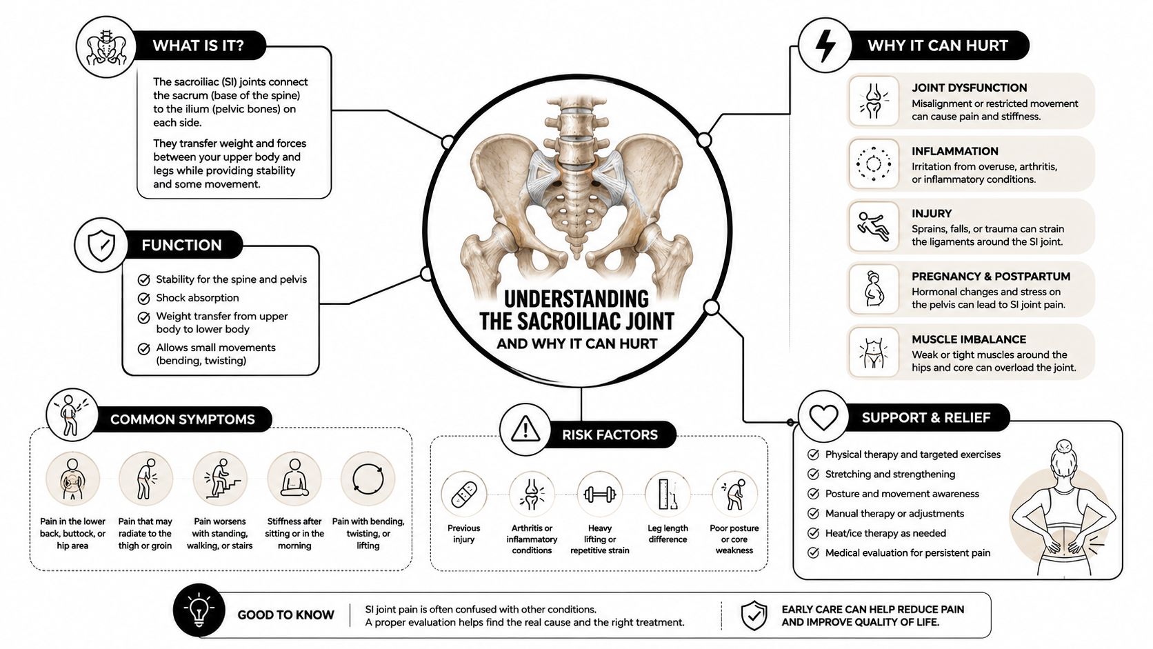
You have two sacroiliac joints, one on each side of the pelvis. Each joint connects the sacrum, the triangular bone at the base of the spine, with an ilium, the broad pelvic bone. Together, they help transfer force between your spine and legs.
A useful analogy is a shock absorber between two moving parts. When you walk, climb stairs, lift, or change direction, the SI joint helps distribute force instead of allowing every impact to travel directly through the lumbar spine. Its movement is limited, which gives the pelvis stability, but the joint still has to manage repeated loading throughout the day.
Why a small joint can create confusing symptoms
Strong ligaments surround the SI joint and help hold it in place. Nerves from several spinal levels supply the joint and nearby tissues, so irritation in one area can feel broader than the joint itself. Pain may remain near the dimples at the back of the pelvis, or it may refer toward the buttock, hip, groin, or thigh.
Several situations can increase stress on the joint:
- Pregnancy-related changes: Altered pelvic mechanics and ligament flexibility can change how forces move through the SI region.
- Degenerative arthritis: Wear within the joint can create painful loading during standing, walking, or transitions.
- Prior lumbar fusion: When motion changes in one part of the spine, nearby joints may carry different forces.
- Trauma: A fall, collision, or sudden twisting movement can irritate the joint or its supporting ligaments.
- Asymmetric loading: A leg-length difference, altered gait, or one-sided movement pattern can repeatedly stress one SI joint.
The same pain pattern can come from a lumbar disc, facet joint, hip joint, or surrounding muscles. That overlap explains why a careful examination matters. An injection isn't just aimed at a sore spot. It tests a specific structure within a crowded neighborhood of possible pain sources.
When the SI Joint Is Worth Investigating as the Pain Source
Pain near one side of the back of the pelvis can point toward the SI joint, especially when it does not follow a classic nerve-root pattern. After disc herniation, disc-related pain, and radiculitis are excluded, reviews estimate that SI joint pain accounts for about 10% to 25% of persistent axial low back pain (review of diagnostic studies). The range is wide because studies use different patient groups and diagnostic methods. It still shows that the SI joint is a reasonable possibility, not a rare explanation.
Suspicion rises when several clues fit together. These may include tenderness near the posterior superior iliac spine, pain when standing from a chair, discomfort during prolonged sitting, symptoms on stairs, or pain referred toward the groin. Symptoms that began after pregnancy, trauma, or lumbar fusion can also make the joint more relevant during the evaluation.
No single symptom confirms the source. Your physician may combine several pain-provocation maneuvers with your history, prior imaging, and an examination of the lumbar spine and hip. The goal is to compare competing explanations rather than judge the SI joint in isolation.
| Pain Source | Typical Location | Common Aggravators |
|---|---|---|
| SI joint | One-sided low back, buttock, pelvis, or groin | Sit-to-stand, stairs, prolonged sitting, standing, turning |
| Lumbar disc | Low back, sometimes into the leg | Sitting, bending, lifting, coughing, or straining |
| Facet joint | More central or one-sided low back | Extension, rotation, standing, and walking |
| Hip joint | Groin, front of the hip, or thigh | Walking, pivoting, hip rotation, and weight bearing |
A systematic evaluation found a point prevalence near 25% in studied populations. It also noted that uncontrolled blocks can produce a false-positive rate of about 20% (review of diagnostic studies). This is why an image-guided injection has two roles: it may offer relief, while the change in your familiar pain provides evidence about whether the SI joint is involved. Your physician must interpret that response alongside the examination and history.
How a Sacroiliac Joint Injection Can Test and Treat at the Same Time
Suppose your familiar buttock pain eases soon after the injection, then returns as the numbing medicine wears off. That short-lived change is not a failed treatment. It is diagnostic information. An SI joint injection combines a diagnostic test with a therapeutic opportunity, so the timing and type of relief matter as much as the relief itself.
The local anesthetic works quickly and temporarily. A clear reduction in your usual pain during its expected window supports the SI joint as a possible source. The corticosteroid has a different role. It aims to calm inflammation and irritation over a longer period, so its effect may build gradually. Do not judge both medicines by the same clock.
Reading the diagnostic response correctly
A single, uncontrolled injection has limits. Pain may improve because of the anesthetic, rest, expectation, or medicine spreading to nearby tissues. For that reason, the response is evidence, not a stand-alone diagnosis. A controlled diagnostic block review explains why clinicians interpret temporary relief alongside your history and examination.
Some clinicians use comparative blocks on separate occasions, with different local anesthetics, to make the conclusion more reliable. Recent consensus guidance generally considers a diagnostic or prognostic block positive at 50% or greater pain relief. Some coverage policies require at least 75% relief on two separate occasions during the expected anesthetic duration (diagnostic coverage policy). Your physician and payer may apply different criteria.
Tracking both parts of the response
Record your pain before the procedure, during the first several hours, and over the following days. Note which activity changed, such as sitting, walking, or rising from a chair. This SI joint pain guide for clinics offers additional background, and an explanation of how a steroid injection works describes the medication's longer anti-inflammatory role.
The most useful follow-up question is specific: Which pain changed, how quickly did it change, and how long did the change last?
What Happens During the Procedure Step by Step
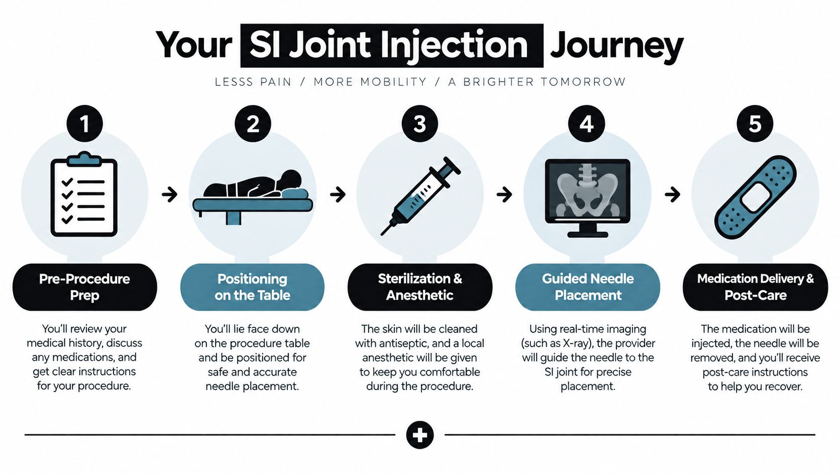
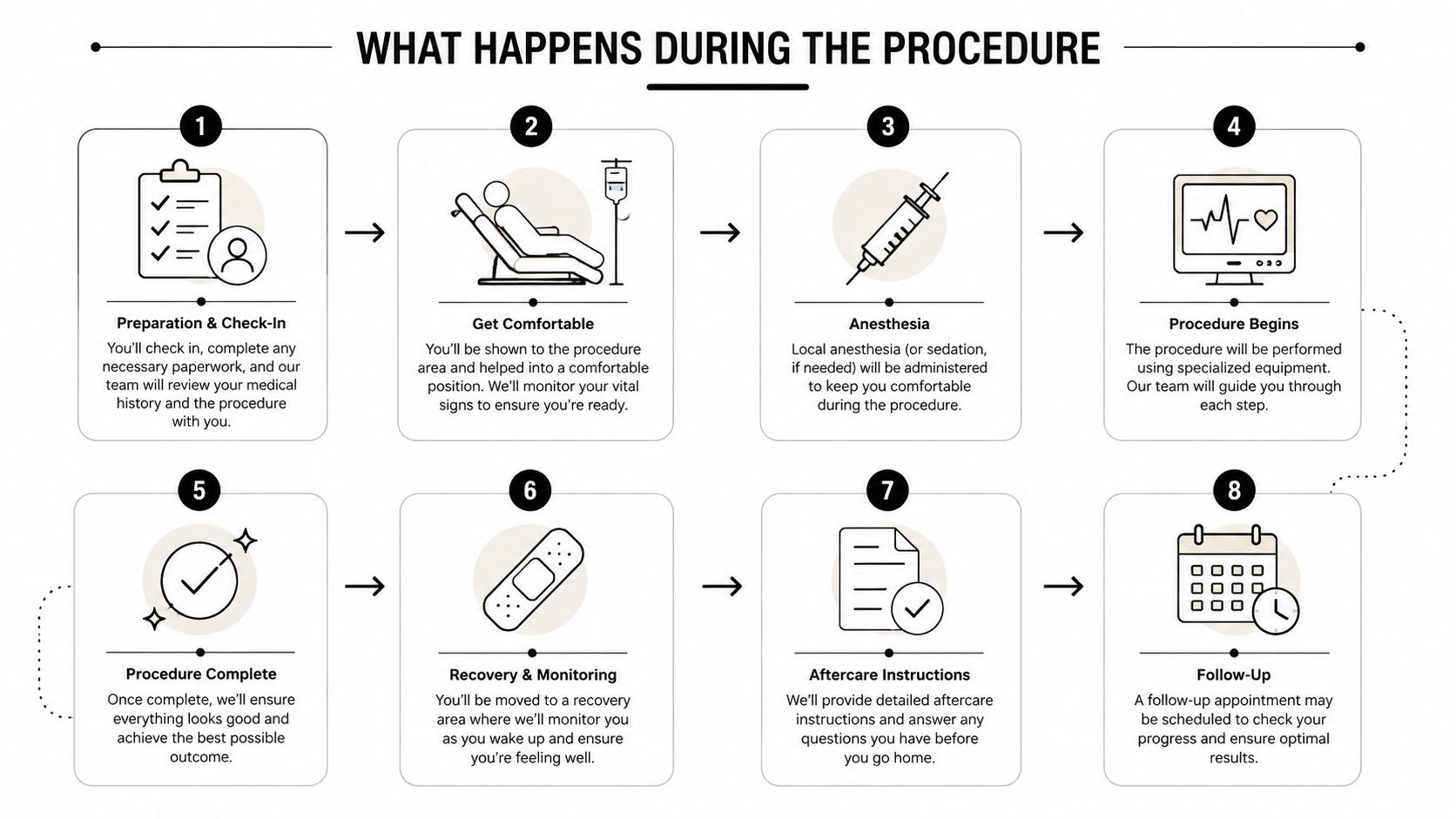
Before the injection, you review your symptoms, medications, allergies, imaging, and the goal of the procedure. Your clinician explains how the same injection may both test the SI joint and calm inflammation, answers questions, obtains consent, and checks vital signs. Blood thinners, diabetes, and other medical conditions may change preparation, so address them before entering the procedure room.
You lie face down on the table, often with support beneath your hips. The skin is cleaned with antiseptic and covered with sterile drapes. Local anesthetic numbs a small patch of skin, sometimes forming a raised wheal, before the deeper needle advances.
Image guidance confirms the target
Fluoroscopy, or live X-ray imaging, guides the needle toward the distal posterior SI joint. Medicare coverage guidance specifies CT or fluoroscopy with contrast for SI joint injections. Ultrasound may be considered in limited situations, such as a documented contrast allergy or pregnancy, although it is less accurate than fluoroscopy for this procedure.
The physician first injects a small amount of contrast. It should outline the joint space and track along the joint line, like dye showing whether a needle has reached the intended narrow compartment. A technical procedure description includes a 22-gauge spinal needle and contrast volumes around 0.3 to 0.5 mL for confirmation in some techniques.
After placement is confirmed, the clinician delivers the medication. The total injectate stays limited. Consensus injection-volume guidance recommends no more than 2.5 mL for an intra-articular SI injection to reduce the chance of capsular rupture and unintended spread. The injection may stop sooner if resistance increases. Pressure or fullness is possible, while sharp pain should be reported immediately.
The procedure commonly takes about 15 to 30 minutes from preparation through completion. Afterward, the team observes you before you stand and walk. Mild soreness or temporary numbness can occur, and light activity usually resumes according to your clinician's instructions. This fluoroscopy-guided injection explanation shows why imaging helps place medication accurately, which matters when the injection is also being used to judge whether the SI joint is contributing to your pain.
Benefits, Success Rates, and What Counts as a Good Response
The main value of an SI joint injection is its two-part role. It can reduce pain for a period of time, while the response also helps clarify whether the SI joint is contributing to your symptoms. That information can guide rehabilitation and later treatment choices.
A practice audit of 155 patients found a positive response after the first injection in 77% of cases. Among 118 responders who received repeat treatment, the mean number of injections was 2.7, and the average response lasted 9.3 months per injection for those needing repeat care. These results describe one clinical setting, not a promise for every patient.
A separate study of 31 patients reported pain relief in 85% at 4 weeks and 74% at 6 months. A newer evidence review found only fair, Level III evidence for therapeutic SI joint injections, based on 11 randomized trials, with 5 positive and 6 negative. The practical lesson is simple: an injection may help, but its result must be judged against your own pain pattern and function.
Function matters as much as the pain score
A useful response might let you sit through a meal, rise from a chair, climb stairs, or complete rehabilitation with less guarding. Pain scores still matter, yet easier movement often shows whether the treatment is helping in daily life.
| Outcome Measure | What a Good Response Looks Like |
|---|---|
| Diagnostic relief | Your familiar SI-region pain decreases during the expected anesthetic window |
| Daily activity | Sitting, walking, standing, or changing position becomes easier |
| Rehabilitation | You can perform core, pelvic, and gluteal exercises that pain previously limited |
| Durability | Relief continues beyond the immediate numbing period and follows a useful pattern |
| Treatment planning | The response helps your physician decide whether to repeat treatment or consider another option |
A response is easier to interpret when it matches the pain that usually troubles you. Provocative examination findings, imaging that supports inflammation, and a prior response to a diagnostic block may strengthen the case for treatment. Injections generally work best as a bridge to movement, posture training, and targeted strengthening, rather than as a stand-alone cure.
If relief fades, the pattern matters. A repeat injection may be reasonable for some patients. Others may discuss radiofrequency ablation of sensory nerves or, after careful evaluation, SI joint fusion. Evidence from a review suggests radiofrequency ablation may help for 3 months to 1 year, although the evidence quality remains low (evidence review of therapeutic injections).
Risks, Side Effects, and Alternatives Worth Knowing
After the injection, the treated area may feel sore. Brief numbness from the anesthetic, flushing, or sleep changes after corticosteroid exposure can also occur. These effects are usually temporary, but tell your clinician if they persist or worry you.
Serious problems such as infection, bleeding, or an allergic reaction are uncommon. Seek medical advice promptly for fever, increasing redness, progressive weakness, or severe pain that keeps worsening. New bowel or bladder changes need urgent medical evaluation.

When relief is incomplete
A brief response still provides information. It may show that the SI joint contributes to your pain while another source also remains active, or that inflammation is not the main driver. Your physician may then reassess the lumbar spine, hip, nerves, gait, and pelvic mechanics.
Treatment choices can include:
- Stability-focused rehabilitation: Exercises may address the abdominal wall, gluteal muscles, pelvic control, and lumbar movement patterns.
- Medication management: Oral anti-inflammatory medication may suit some patients, depending on kidney function, stomach health, cardiovascular risk, and other conditions.
- Radiofrequency ablation: This procedure targets sensory nerves around the SI joint and may be considered when diagnostic blocks support that pain pathway.
- Regenerative options: Platelet-rich plasma and cell-based treatments remain areas of clinical use and investigation. Suitability depends on the patient and treatment setting.
- Surgical consultation: SI joint fusion is more invasive and reserved for selected cases involving persistent, well-localized pain despite other care.
Information about conservative back pain care can help you review nonoperative approaches. Your clinician can also discuss SI joint pain treatment options alongside your examination and injection response. These options are not a fixed ladder. The injection's diagnostic result helps show whether the SI joint deserves further treatment, or whether another pain source needs attention.
Working With the Midwest Pain & Wellness Team Next
A productive consultation starts with useful details, not a perfect explanation. Bring any relevant spine, pelvis, or hip imaging reports, a current medication list, records from prior injections, and notes about treatments that helped or failed. If you have images on a disc or through a patient portal, ask how the clinic prefers to receive them.
Keep a short pain diary before the visit. Note whether symptoms worsen with sit-to-stand movement, climbing stairs, prolonged sitting, walking, rolling in bed, or standing on one leg. Also record whether pain stays in the buttock or travels toward the groin, hip, or leg.
Questions that clarify the plan
Ask the physician:
- Diagnostic reasoning: How will you distinguish SI joint pain from disc, facet, hip, or nerve pain?
- Imaging technique: Will fluoroscopy or another image-guided method be used, and will contrast confirm placement?
- Medication purpose: Which part of the injection is intended to test the joint, and which part is intended to reduce inflammation?
- Response criteria: What degree and timing of relief would count as meaningful?
- Next steps: If relief is brief, would rehabilitation, repeat treatment, radiofrequency ablation, or another evaluation make sense?
Midwest Pain & Wellness is a pain and wellness clinic in Chicago Ridge, Illinois, serving nearby communities including Palos Heights, Palos Hills, Evergreen Park, and Hickory Hills. Its interventional pain team can evaluate whether SI joint pain fits your history and examination, coordinate with primary care or other clinicians when appropriate, and use diagnostic findings to shape a broader plan that may include physical therapy or radiofrequency ablation.
Bring your questions, describe the pain in everyday terms, and ask for a timeline that matches the uncertainty of diagnosis. A careful evaluation can turn a frustrating pain pattern into a clearer set of decisions.
Midwest Pain & Wellness offers evaluation and image-guided sacroiliac joint injections for patients with persistent low back or buttock pain in the Chicago Ridge area. Visit Midwest Pain & Wellness to learn more and schedule a consultation about whether an SI joint injection fits your symptoms and treatment goals.