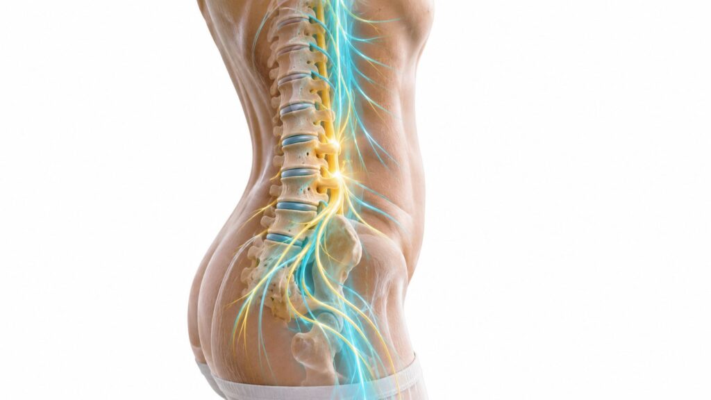
Persistent pain low in the back or deep in the buttock can make ordinary movements feel strangely difficult. Standing from a chair, climbing stairs, turning in bed, or walking through a grocery store may all irritate the same small area beside the sacrum. If treatment, medication, and activity changes haven't settled the problem, you may be wondering whether the sacroiliac joint is involved and what an SI joint injection technique involves.
An injection is not just a needle placed near the painful spot. The clinician uses the procedure to answer a diagnostic question, deliver medication to a specific joint, and decide what treatment makes sense next. For patients in Chicago Ridge, Palos Heights, Palos Hills, Evergreen Park, and Hickory Hills, Illinois, understanding the reasoning behind each technical choice can make the appointment less intimidating and the results more useful.
Table of Contents
- When an SI Joint Injection Technique Makes Sense
- Why Imaging Guidance Changes Everything
- Getting Ready for the Procedure
- What Happens During the Injection
- Diagnostic Value and What the Results Tell Us
- Risks, Side Effects, and Recovery
- After the Injection and Long-Term Planning
When an SI Joint Injection Technique Makes Sense
Finding the likely pain generator
SI joint pain often sits near the posterior superior iliac spine, the bony area commonly described as the “dimple” beside the lower back. It may remain in the buttock or extend toward the groin or thigh. That pattern can resemble pain from the lumbar discs, facet joints, hip, or surrounding muscles, so location alone doesn't establish the diagnosis.
A pain specialist looks for a consistent history and examines how the pelvis responds to controlled stress. The Fortin finger test asks where the patient points when identifying the worst pain. Other maneuvers include Patrick's FABER test, thigh thrust, pelvic compression, and distraction. When several of these tests reproduce the familiar pain, suspicion increases that the SI joint may be contributing to the symptoms.
An injection becomes more reasonable when the pain has persisted despite measures such as physical therapy, oral medication, and activity modification. It may also be considered in degenerative sacroiliitis, pain after trauma, inflammatory spondyloarthropathy, or symptoms that develop after lumbar fusion. None of those conditions automatically proves that the SI joint is the source, however.
Practical rule: An examination can raise suspicion, but a carefully performed anesthetic injection provides more direct information about whether the joint is generating the familiar pain.
One procedure, two purposes
The diagnostic portion uses local anesthetic to temporarily interrupt pain from inside the joint. If the patient's usual pain is reproduced during needle placement or medication delivery and then meaningfully improves during the anesthetic window, the result supports the SI joint as a pain generator.
The therapeutic portion uses an anti-inflammatory medication, commonly a corticosteroid, to calm irritation within the joint. Relief can be brief, prolonged, or absent depending on the underlying pathology. A technically successful injection won't correct pain that originates in the lumbar spine, hip, peripheral nerves, or another structure.
That distinction matters. The procedure isn't a guarantee of permanent relief, and a short response isn't automatically a failure. It may confirm the joint's involvement while showing that a different long-term strategy is needed.
Why Imaging Guidance Changes Everything
A patient can lie face down, remain still, and still receive an injection that misses the SI joint. Landmark-only technique relies on surface anatomy and the clinician's estimate of the joint's position beneath the tissues. In a large review, intra-articular placement without imaging succeeded in only 22% of cases. Epidural flow occurred in 24%, and dorsal sacral foraminal flow in 44%, showing how easily medication can enter nearby structures instead. The same review of image-guided sacroiliac joint injections reported fluoroscopy-guided accuracy of up to 97% with appropriate confirmation.
The anatomy creates the challenge. The SI joint is deep, narrow, and angled, not a flat target directly beneath the skin. Its posterior recess is small, with sacral foramina and neural structures nearby. A needle may feel correctly positioned while remaining outside the capsule. That matters clinically because misplaced anesthetic can produce a false diagnostic result, and misplaced steroid may treat the wrong tissue.
Comparing the guidance choices
| Guidance method | Intra-articular accuracy | Radiation exposure |
|---|---|---|
| Blind landmark technique | 22% in a large review | None |
| Fluoroscopy with contrast confirmation | Up to 97%, large review, PMC7041665 | Yes |
| Fluoroscopy versus ultrasound | 98.2% fluoroscopy, prospective randomized trial, Pain Medicine 2019 | Yes |
| Ultrasound | Lower accuracy than fluoroscopy in the cited comparison | None |
The 98.2% fluoroscopy accuracy figure comes from a prospective randomized comparison reported in published SI joint injection research. It describes a different study design from the review reporting up to 97% with contrast confirmation, so the figures should not be read as competing measurements of one procedure. CMS guidance also cites 98% accuracy with fluoroscopy versus 87% with ultrasound and generally calls for CT or fluoroscopy with contrast, with limited exceptions such as documented contrast allergy or pregnancy. The CMS coverage guidance for SI joint injections provides that policy context.
Fluoroscopy shows bony landmarks, helps align an oblique view with the joint cleft, and confirms spread after a small amount of contrast. The trade-off is ionizing radiation. Ultrasound avoids radiation and can display soft tissue and vessels dynamically, but deeper tissue or unusual anatomy may make the joint harder to identify.
In a community pain clinic serving the Chicago Ridge area, the guidance choice should match the clinical question, anatomy, and safety needs. Injectate volume also matters. Too much fluid can spread beyond the joint and make a diagnostic response harder to interpret. Patients can review what a fluoroscopy-guided injection involves before discussing the approach with the clinician.
Getting Ready for the Procedure
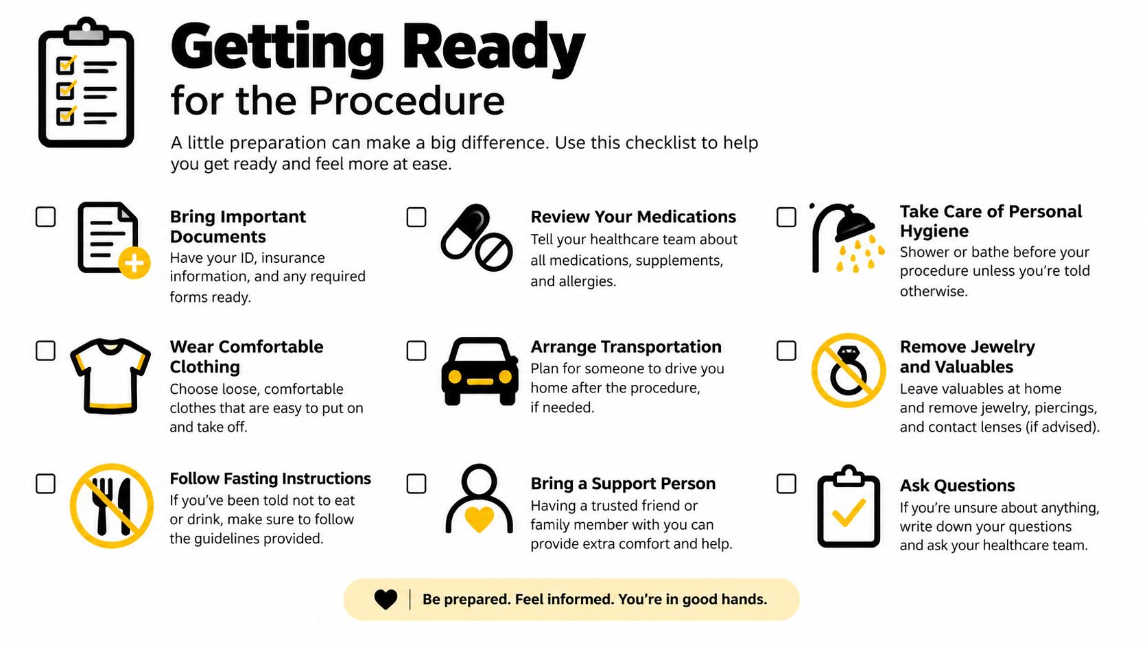
Preparation starts with the consultation, not the procedure table. The physician reviews your symptoms, prior imaging, medical history, current medications, and previous injections or operations. Tell the clinical team about anticoagulants, antiplatelet medication, bleeding disorders, diabetes, pregnancy, and allergies to iodinated contrast, local anesthetic, or steroid medication.
Medication instructions must be individualized. Don't stop a blood thinner or antiplatelet drug on your own. The prescribing clinician and interventional pain specialist decide whether a temporary change is safe, based on why you take the medication and the bleeding risk of the planned procedure.
Questions to settle beforehand
The consent conversation should cover the expected diagnostic value, possible relief, alternatives, and risks. It should also clarify whether sedation is planned, whether you need to fast, and whether someone must drive you home. Requirements vary by clinic and by the medications used, so follow the instructions provided for your appointment rather than relying on a general internet checklist.
Wear clothing that allows access to the lower back and pelvis. Remove items that could interfere with positioning, and tell the staff if lying face down is difficult because of breathing problems, shoulder pain, reflux, or another condition.
Positioning and skin preparation
In the procedure room, you'll lie prone on a firm imaging table. A small support under the pelvis may reduce the lumbar curve and make the posterior SI joint easier to access. The C-arm is then adjusted obliquely so the clinician can see the inferior portion of the joint more clearly.
The skin is cleaned with an antiseptic such as chlorhexidine or povidone-iodine and covered with sterile drapes. Local anesthetic, often lidocaine, numbs the skin and tissues along the needle path. Expect a brief sting or burning sensation from the anesthetic, followed by pressure as the clinician works. The table can feel firm, so tell the team if you need a position adjustment before the sterile field is established.
What Happens During the Injection
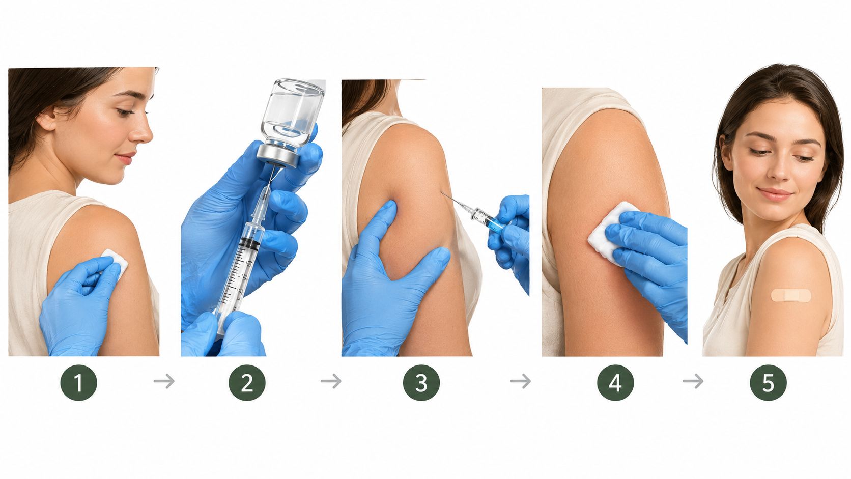
Once positioning and skin preparation are complete, the needle portion begins with you face down and the lower back supported in a stable position. Under fluoroscopy, the clinician adjusts the C-arm to open the posterior SI joint cleft rather than viewing the pelvis straight on. This oblique view helps identify the inferior joint and guides the entry point a few centimeters lateral to the sacral midline.
A small wheal of local anesthetic numbs the skin. The physician then advances a fine, commonly 22- or 25-gauge needle along a controlled coaxial path toward the inferior pole of the joint. Pressure, pushing, or brief reproduction of your familiar pain can occur. That feedback may help with orientation, while sharp or escalating pain should be reported immediately.
Why contrast confirmation matters
The needle must be confirmed inside the joint before medication is delivered. Under live fluoroscopy, the clinician injects only 0.3 to 0.5 mL of contrast to observe its spread and verify the needle tip. The consensus guidance on sacroiliac joint complex pain addresses this technical step and the limits on injectate volume. A satisfactory arthrogram shows contrast tracking within the intended joint space.
The contrast pattern can change the plan immediately. Spread outside the capsule suggests extra-articular placement. Rapid vascular uptake raises concern that the needle is in or near a blood vessel. The clinician may reposition the needle, obtain another view, or stop rather than assume the medication will reach the target.
The final injectate combines local anesthetic with an anti-inflammatory medication in a deliberately small volume. Volume control matters because excess fluid can rupture the capsule or spread into nearby structures, making the result harder to interpret. The 2024 multispecialty consensus guideline recommends a maximum total injectate volume of 2.5 mL. That limit helps preserve the diagnostic value of the injection while reducing unwanted spread.
Stay still, but report what you feel. Pressure, burning, familiar pain, and a new sharp sensation mean different things during needle placement. After the joint is confirmed and the medication is delivered, the needle is withdrawn, the site is covered, and the team observes you according to the clinic's routine.
Diagnostic Value and What the Results Tell Us
The most useful question isn't just, “Did my pain improve?” The clinician also wants to know whether the injection reproduced your familiar pain, how much that pain changed during the anesthetic phase, which activities became easier, and whether the improvement matched the expected duration of the local anesthetic.
A strong response supports the diagnosis, particularly when the pain that appears during the procedure is the same pain you experience at home. A technically accurate injection that doesn't reduce the familiar symptoms points the evaluation elsewhere, possibly toward lumbar facet joints, a disc, the hip, or another pain source. A negative result can be valuable because it prevents repeated treatment aimed at the wrong structure.
How clinicians interpret the response
| Response pattern | Diagnostic implication | Typical next step |
|---|---|---|
| Familiar pain is reproduced, then substantially improves during the anesthetic phase | Supports the SI joint as a pain generator | Track function and discuss treatment directed at the SI joint |
| Relief is partial or difficult to separate from normal fluctuation | The SI joint may contribute, but another source may also be active | Reassess the examination, pain pattern, and competing diagnoses |
| No meaningful change after technically confirmed placement | Makes the SI joint a less likely primary source | Evaluate the lumbar spine, hip, nerves, or other structures |
| Relief occurs only after the anti-inflammatory phase | Suggests a therapeutic response, but may provide less immediate diagnostic clarity | Record duration and functional improvement before deciding on further care |
Some clinicians and payers use dual comparative blocks, meaning different local anesthetics are used on separate visits to reduce the chance that expectation or a nonspecific response will determine the diagnosis. Requirements vary, so patients should ask what threshold the treating clinician and insurer use.
Volume is part of interpretation. If medication spreads beyond the target, it can numb nearby posterior sacral nerve branches and create an apparent improvement that doesn't come solely from the SI joint. This is why accurate needle placement and restrained injectate volume matter as much as the medication itself. The broader difference between anti-inflammatory treatment and diagnostic anesthetic response is explained in this overview of how steroid injections work.
Risks, Side Effects, and Recovery
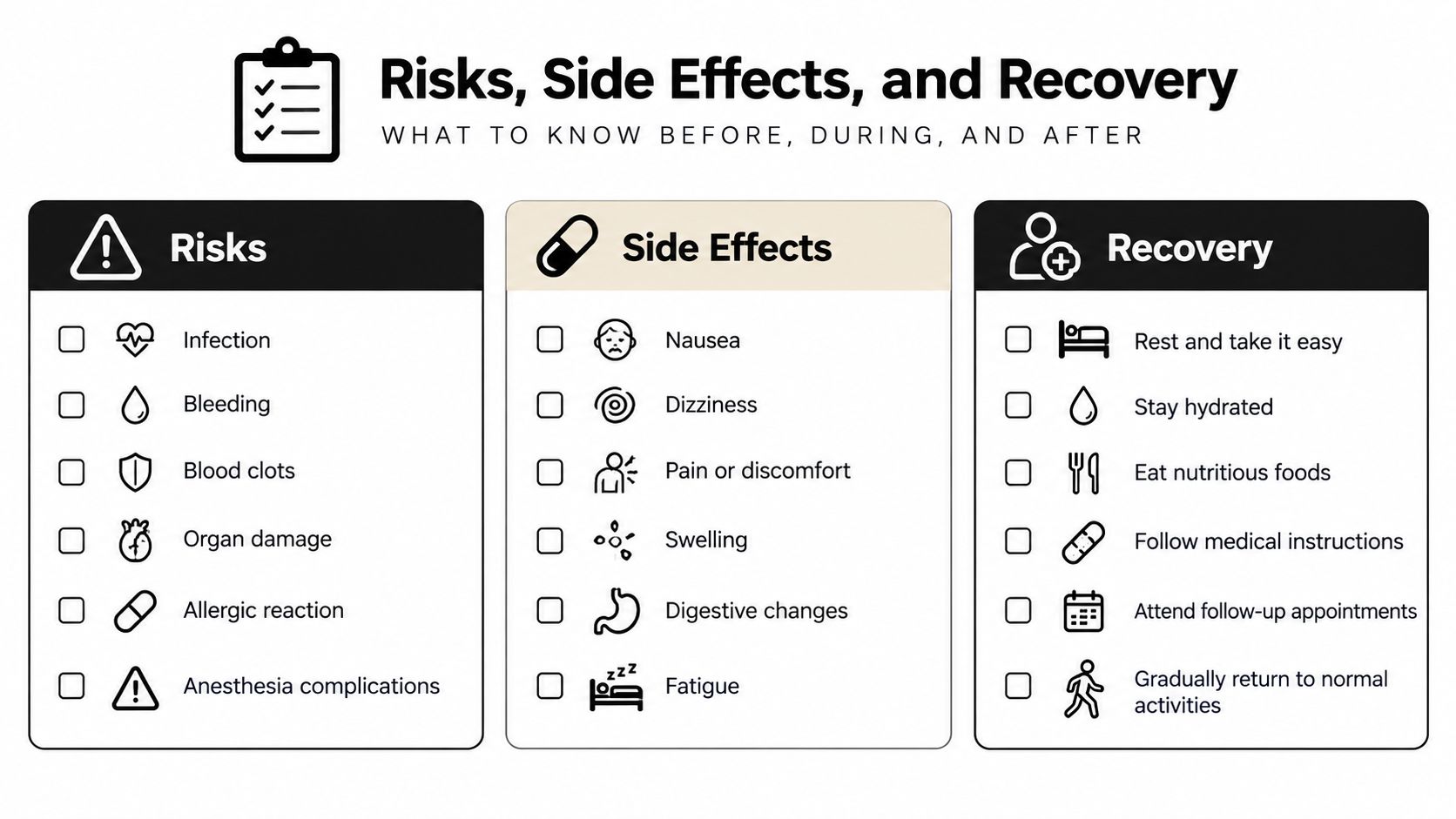
Image guidance improves precision, but it doesn't eliminate risk. Bleeding, infection, contrast or medication reaction, and temporary soreness can occur with any injection. More specific complications include nerve irritation or injury, unintended nerve root block, hematoma, vascular uptake of medication, and systemic effects from corticosteroid treatment. Clinical summaries describe most adverse effects as minor while still recognizing these less common serious complications in the clinical reference on SI joint injections.
The technical choices reduce rather than erase those concerns. Sterile preparation limits contamination. Contrast under live imaging helps identify an unwanted vascular or extra-articular pattern before the therapeutic medication is delivered. A small total volume reduces the chance of pressure-related capsular rupture and spread to neighboring structures.
What recovery usually feels like
Mild soreness at the entry site, temporary numbness, or a feeling of heaviness can occur after the anesthetic. Some people experience a corticosteroid flare, in which the treated area feels more painful before it settles. Avoid judging the final result while the local anesthetic is active or while a temporary flare is developing.
Most patients can return to gentle daily movement according to the clinician's instructions. Avoid strenuous activity until the team says it is appropriate, then resume normal movement gradually. A pain diary can distinguish procedure soreness from the original pain pattern.
Contact the clinic promptly for fever, increasing redness or swelling, severe headache, new weakness, or changes in bowel or bladder control. Those symptoms aren't routine post-injection soreness and deserve direct medical assessment rather than watchful waiting.
After the Injection and Long-Term Planning
The anesthetic response may appear quickly and then fade, while anti-inflammatory benefit develops more gradually. Some patients notice improvement within 24 to 48 hours, and corticosteroid benefit commonly develops over 3 to 7 days, but individual timing varies. Don't decide that the procedure failed during the first uncomfortable hours.
Record pain intensity, walking or standing tolerance, sleep, medication use, and the activities that trigger symptoms. Also write down whether the injection relieved the familiar pain or only created a general sense of numbness. Those details help the clinician determine whether the SI joint was confirmed as a pain source and whether the response was useful enough to guide the next decision.
Using the result to choose the next treatment
Relief that lasts weeks to several months can support a plan directed at the SI joint. Depending on the response and clinical findings, that plan may include radiofrequency neurotomy of the lateral sacral branches for longer-term control. Brief relief can still be diagnostically meaningful, but it may also prompt a closer search for lumbar facet, discogenic, hip, or nerve-related pain.
An injection should function as a checkpoint, not the entire care plan. Physical therapy may address pelvic asymmetry, sacral belt stabilization, movement coordination, and progressive core strengthening. The goal is to improve standing, walking, sleep, and daily function while limiting unnecessary repeat steroid exposure.
At a community pain clinic serving Chicago Ridge and nearby Illinois communities, reassessment typically focuses on the diary, examination, functional change, and the duration of benefit. The discussion may lead toward another injection when appropriate, denervation, rehabilitation, or evaluation of another pain generator. Patients can review broader sacroiliac joint pain treatment options before that follow-up conversation.
Midwest Pain & Wellness offers image-guided SI joint injections, diagnostic evaluation, and coordinated pain care for patients in the Chicago Ridge area. Visit Midwest Pain & Wellness to discuss whether an SI joint injection technique is appropriate for your symptoms and to plan care around lasting function rather than temporary relief alone.