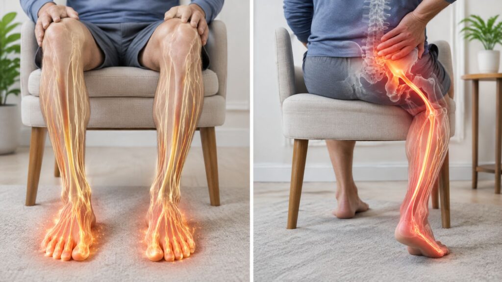
You've been waking up with tingling in your hand, or perhaps pain has started traveling from your neck into your arm or from your lower back into your leg. Rest helps briefly, but the symptoms return when you type, walk, sleep, or sit. The question usually isn't, “Do I need surgery?” It's, “Which nerve is irritated, where is it being compressed, and what treatment fits that exact problem?”
Nerve compression treatment works best as a diagnosis-specific plan. A wrist entrapment, a cervical disc problem, lumbar spinal stenosis, and a nerve compressed near the elbow may all produce numbness or weakness, but they don't share the same anatomy or the same treatment pathway. This guide explains how specialists distinguish those conditions, what conservative care can accomplish, when image-guided procedures make sense, and why timing matters.
Table of Contents
- What Nerve Compression Really Feels Like
- How Nerves Get Compressed in the First Place
- Symptoms and How Specialists Confirm the Diagnosis
- Conservative Care That Comes Before Any Procedure
- Image-Guided Injections, Nerve Blocks, and Ablation
- Stimulators, Minimally Invasive Decompression, and Regenerative Options
- Why Timing and the Right Diagnosis Change the Outcome
- When to Seek Specialist Care and What to Expect Next
What Nerve Compression Really Feels Like
A 52-year-old office worker once described months of nighttime hand tingling as “shoulder tightness.” She adjusted her chair, stretched her neck, and blamed long hours at a computer. During an examination, the pattern told a different story. The symptoms traveled from the neck into the arm and hand, and further evaluation identified cervical nerve root compression related to a herniated disc.
That distinction matters. A compressed nerve isn't a sore muscle. It's a mechanical irritation at a particular point along the nerve's route. The irritated nerve can send abnormal signals far from the original pressure, which is why a problem in the neck may be felt in the hand, and a problem in the lower back may be felt in the foot.
The sensations patients commonly describe
People use different words for nerve symptoms, but the underlying pattern is often recognizable:
- Pins and needles: A prickling or “asleep” feeling that may come and go.
- Burning: Heat or a raw sensation that doesn't match what the skin looks like.
- Electric shocks: Brief jolts triggered by movement, coughing, or certain positions.
- Deep aching: A persistent discomfort that follows an arm or leg rather than staying in one joint.
- Weak grip or foot control: Difficulty opening a jar, holding objects, lifting the front of the foot, or climbing stairs.
- Reduced sensation: Skin may feel numb, dull, or different when touched.
Muscle pain usually stays closer to the muscle and often changes with direct use or pressure. Joint pain commonly centers around a specific joint and may be tied to movement, swelling, or stiffness. Nerve pain tends to travel along a recognizable territory, and it may combine pain with tingling, altered sensation, weakness, or changes in reflexes.
Practical rule: Pain location isn't always the same as compression location. The nerve's pathway often provides a more useful clue than the place that hurts most.
Carpal tunnel syndrome illustrates how common these problems can be. It's the most common peripheral nerve compression syndrome, with lifetime prevalence estimated at about 10% in one review and prevalence placed at roughly 3% in the general population and about 15% in the workforce in another clinical review (clinical review of carpal tunnel syndrome). Those figures describe different populations and methods, so they shouldn't be treated as contradictory.
The evaluation that follows should connect your symptoms to anatomy. A pain specialist considers the nerve territory, strength, reflexes, provocative movements, imaging, and sometimes nerve studies before recommending nerve compression treatment. The sections ahead move from anatomy and diagnosis through conservative care, image-guided procedures, advanced options, timing, and the warning signs that call for prompt specialist attention.
How Nerves Get Compressed in the First Place
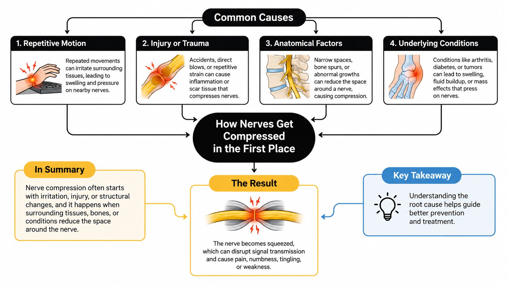
A nerve becomes compressed when surrounding tissue reduces the space available for it or applies pressure at a vulnerable anatomical site. The same phrase, “pinched nerve,” can therefore describe very different conditions. Treatment depends on whether the pressure occurs at a spinal nerve root, a peripheral nerve tunnel, or a region where several structures pass through a confined space.
Location determines the problem
In the cervical spine, a herniated disc or foraminal narrowing can crowd a nerve root as it exits the spine. In the lumbar spine, central canal or lateral recess narrowing can reduce room for nerves traveling toward the legs. Spinal stenosis is a structural diagnosis, not just a description of back pain.
The thoracic outlet is another distinct region. The brachial plexus and nearby subclavian vessels pass between the clavicle and first rib, so compression there can produce arm symptoms that don't follow the same pattern as carpal tunnel or cervical radiculopathy.
Peripheral nerves can also become trapped in narrow passageways:
- Carpal tunnel: The median nerve is compressed at the wrist.
- Cubital tunnel: The ulnar nerve is irritated near the elbow.
- Peroneal nerve region: The common peroneal nerve can be compressed near the fibular head.
- Tarsal tunnel: Structures around the ankle can crowd the tibial nerve.
What creates pressure
Bone spurs, thickened ligaments, herniated disc material, swollen tendons, cysts, tumors, trauma, and inflammation can all reduce space around a nerve. Repetitive positioning or direct pressure may worsen symptoms, but the underlying anatomy still needs to be identified before treatment is selected.
Sustained pressure can interfere with blood flow inside the nerve. Over time, that stress can affect the insulating myelin layer and, with more prolonged injury, the nerve fibers themselves. The result may progress from intermittent tingling to persistent numbness or weakness.
The double-crush concept adds another layer. A nerve already irritated at the neck may tolerate less pressure at the wrist, so a smaller downstream entrapment can contribute to symptoms. Metabolic conditions, including diabetes, hypothyroidism, and obesity, may further increase nerve vulnerability. That's why a careful assessment looks beyond the single spot that hurts.
Symptoms and How Specialists Confirm the Diagnosis
Symptoms become more useful when you map them to a nerve territory. Cervical radiculopathy may travel into the shoulder, arm, or hand. Lumbar radiculopathy may extend from the back into the buttock, leg, or foot. Thoracic outlet symptoms can involve the ulnar side of the arm, while carpal tunnel commonly affects the thumb, index finger, middle finger, and part of the ring finger. Cubital tunnel symptoms more often involve the ring and small fingers.
| Compression Site | Typical Symptom Pattern |
|---|---|
| Cervical nerve root | Neck pain with symptoms into the shoulder, arm, or hand |
| Lumbar nerve root | Low-back pain with symptoms into the buttock, leg, or foot |
| Thoracic outlet | Arm symptoms, often toward the ulnar side, with symptoms affected by shoulder position |
| Carpal tunnel | Tingling or numbness in the thumb, index, middle, and part of the ring finger |
| Cubital tunnel | Tingling or numbness in the ring and small fingers |
| Peroneal nerve near the fibular head | Sensory symptoms over the outer lower leg or top of the foot, sometimes with weakness lifting the foot |
What the exam looks for
A pain specialist begins with the history. The timing of symptoms, aggravating positions, sleep disruption, work demands, injuries, medical conditions, and changes in strength all help narrow the possibilities.
The neurological exam then tests sensation, motor function, reflexes, gait, and specific maneuvers. Depending on the suspected location, the clinician may use Spurling testing for cervical nerve irritation, straight leg raise for lumbar radicular symptoms, Phalen and Tinel testing for carpal tunnel, and Adson testing when thoracic outlet compression is under consideration.
Imaging answers different questions. MRI provides detail about discs, nerves, and other soft tissues. CT shows bony anatomy in greater detail. X-rays can help assess alignment and instability. Ultrasound can evaluate accessible peripheral nerves dynamically.
Electromyography and nerve conduction studies help localize the lesion, estimate severity, and distinguish focal compression from a more diffuse peripheral neuropathy. The aim isn't to collect tests for their own sake. It's to establish a precise anatomical diagnosis before choosing nerve compression treatment. For a plain-language overview of other conditions that can produce back and leg symptoms, see this resource on causes of back and leg pain. You can also review the clinical process in this guide to how nerve pain is diagnosed.
Conservative Care That Comes Before Any Procedure
Many nerve compression problems begin with nonoperative care, but “conservative” doesn't mean passive. The purpose is to reduce mechanical irritation, control inflammation, maintain safe movement, and watch for signs that the nerve isn't recovering.
Cleveland Clinic describes common initial options such as splints, NSAIDs, steroid injections, physical or occupational therapy, with surgery considered when nonsurgical care fails (nerve compression syndrome treatment options). The appropriate combination depends on the site and severity.
Four useful first-line tools
Activity modification reduces the positions that repeatedly load the affected nerve. That might mean changing wrist posture, avoiding prolonged elbow flexion, limiting heavy lifting for a period, or adjusting sitting and walking habits. It can calm symptoms, but it usually won't remove a bone spur or enlarge a narrowed canal.
Medication may include an NSAID when inflammation and pain are prominent, or a prescription nerve-pain medication when burning and electrical symptoms dominate. These medicines require attention to kidney function, stomach risk, sedation, interactions, and other health factors. A medication should earn its place by improving sleep, movement, or participation in rehabilitation.
Splinting and bracing place a joint in a less provocative position. A nighttime wrist splint can reduce repeated bending at the wrist, while an elbow strategy may limit prolonged flexion. The brace should support healing without creating new stiffness or weakness.
Physical therapy has two different jobs. It can reduce axial loading through posture, graded strengthening, and movement retraining, while nerve-gliding exercises aim to help the nerve move through surrounding tissues without aggressive stretching. Those are not interchangeable goals.
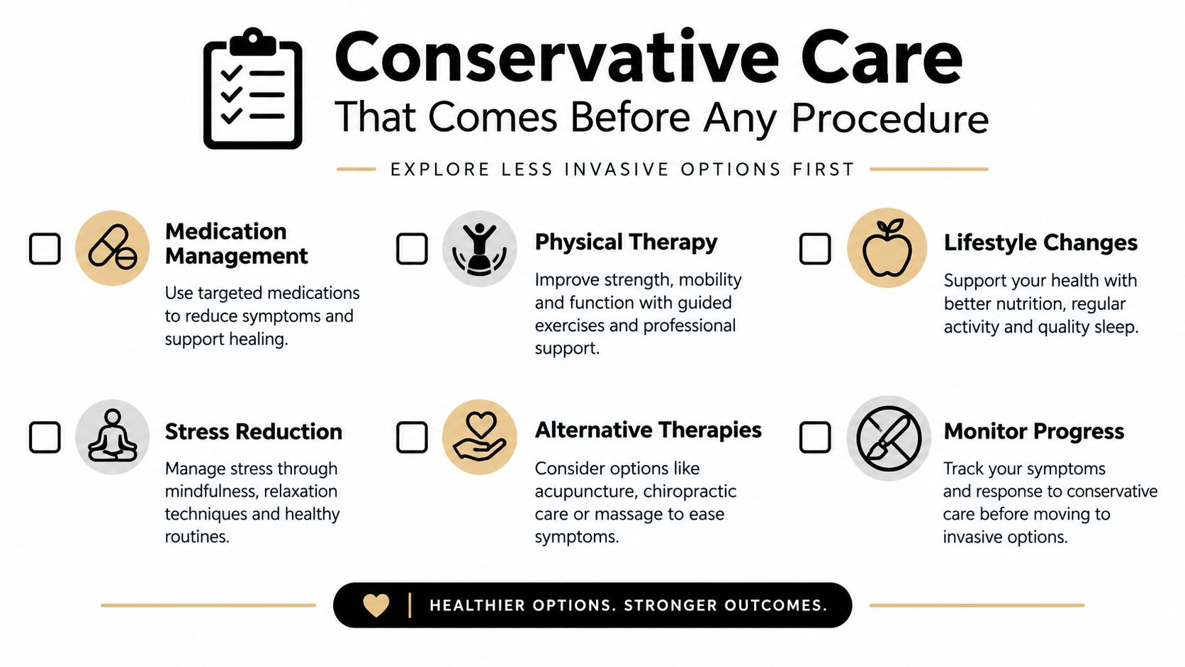
A structured trial often lasts about 6 to 12 weeks, but the calendar doesn't override the examination. Progressive motor weakness should shorten the trial, and bowel or bladder changes require urgent medical evaluation rather than routine observation. A limited conservative plan may also continue alongside a procedure when the intervention is intended to create enough relief for rehabilitation.
Image-Guided Injections, Nerve Blocks, and Ablation
When symptoms plateau but the anatomy doesn't yet call for surgery, image-guided procedures can answer two questions. Can treatment calm the irritated structure, and does a targeted intervention confirm the suspected pain generator?
An epidural steroid injection places medication near an inflamed spinal nerve root under fluoroscopic or ultrasound guidance, depending on the procedure and region. It's most useful when symptoms match a documented nerve-root problem. For epidural injections used in radiculopathy and spinal stenosis, a systematic review found that immediate pain relief favored injections, but the benefit was small and didn't persist at longer follow-up; immediate functional improvement wasn't statistically significant (systematic review of epidural corticosteroid injections). That's why an injection should be presented as a targeted tool, not a guaranteed structural fix.
A selective nerve root block places local anesthetic, sometimes with an anti-inflammatory medication, near a specific exiting nerve. Relief can support the diagnosis and may provide temporary symptom control. For patients trying to understand the procedure in plain language, this explanation of what a nerve block injection does can be useful.
Matching the procedure to the suspected generator
Medial branch blocks target small nerves that carry pain from facet joints. They're diagnostic when the pain pattern and examination suggest facet-mediated pain, not when the primary problem is direct compression of a spinal nerve root. If properly selected blocks provide meaningful confirmation, radiofrequency ablation can use controlled heat to interrupt pain signaling from those nerves.
| Procedure | What It Targets | Typical Duration of Relief |
|---|---|---|
| Epidural steroid injection | Inflamed spinal nerve root | Often temporary, with response varying by diagnosis |
| Selective nerve root block | A specific nerve root for diagnostic and therapeutic purposes | Usually temporary |
| Medial branch block | Nerves supplying painful facet joints | Short-term diagnostic or therapeutic relief |
| Radiofrequency ablation | Confirmed facet-related pain signaling | Longer-lasting relief in appropriate candidates |
Patients often expect injections to last indefinitely. In practice, relief may be temporary, and repeated procedures become less useful if a fixed compression remains untreated. Imaging must also match the examination. A Carelon guideline states that epidural injections for nerve-root compression from a herniated disc require MRI or CT imaging from within the previous 18 months, with imaging findings that correlate with clinical findings (interventional pain management guideline).
Stimulators, Minimally Invasive Decompression, and Regenerative Options
Advanced treatments aren't a single final step. Each one addresses a different problem, and selecting among them requires a clear diagnosis.
Peripheral nerve stimulation can be considered for a defined peripheral nerve pain pattern when the nerve is generating persistent pain but direct decompression isn't the immediate answer. Spinal cord stimulation is used for selected patients with persistent, often more diffuse limb pain. Neither treatment creates more room around a mechanically compressed nerve. Stimulation changes pain signaling, so it shouldn't be mistaken for decompression.
Procedures that create space
Minimally invasive lumbar decompression, including MILD, removes selected tissue through a small access point when ligament-related lumbar spinal stenosis is the relevant anatomy. Interspinous stabilization procedures such as Vertiflex are designed for particular patterns of lumbar stenosis and aren't suitable for every spinal level or every form of narrowing.
Kyphoplasty belongs to a different category. It treats selected vertebral compression fractures, not routine nerve entrapment. That distinction prevents a procedure from being chosen because it sounds minimally invasive rather than because it matches the source of symptoms.
Regenerative options such as platelet-rich plasma and cell-based therapies are also diagnosis-dependent. They may be discussed when tissue irritation or degeneration is part of the clinical picture, but they shouldn't be marketed as a universal way to release a compressed nerve. Evidence and appropriate use vary by condition, target tissue, and technique. A more detailed explanation of platelet-rich plasma benefits can help patients prepare questions for a consultation.
Midwest Pain & Wellness in Chicago Ridge offers coordinated interventional pain care that may include image-guided injections, nerve blocks, radiofrequency ablation, stimulation, minimally invasive lumbar decompression, kyphoplasty, and regenerative options. The relevant question isn't which treatment is newest. It's whether the treatment addresses the actual source of your symptoms.
Why Timing and the Right Diagnosis Change the Outcome
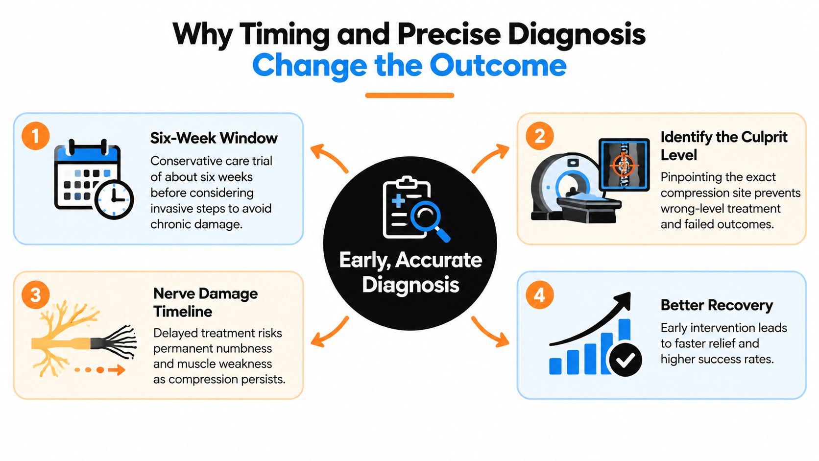
The best outcome often depends less on choosing an impressive procedure and more on identifying the correct nerve and acting before weakness becomes established. A short, structured period of conservative care can be reasonable when symptoms are stable and strength is preserved. It gives the clinician time to see whether irritation settles while the patient works on movement, positioning, and function.
That approach changes when the nerve is losing function. Progressive weakness, worsening gait, or bowel and bladder symptoms can signal a problem that needs urgent evaluation. Persistent compression may move from reversible irritation toward lasting numbness or muscle weakness, so waiting indefinitely for pain alone to improve isn't a safe strategy.
Why one treatment site may not explain everything
Double-crush syndrome can make a single-site plan look unsuccessful. A nerve compressed in the neck may become more sensitive to a second compression at the wrist, and symptoms can persist if a clinician treats only one site. A 2026 systematic review of upper-limb double crush syndrome reported that bimodal decompression, addressing both proximal and distal compression, improved pain and function more than single-site decompression (systematic review of upper-limb double crush syndrome).
This is why persistent numbness after an apparently successful carpal tunnel or ulnar nerve procedure deserves reassessment rather than an automatic conclusion that the procedure failed. The clinician may need to revisit the neck, thoracic outlet, peripheral nerve, or another level.
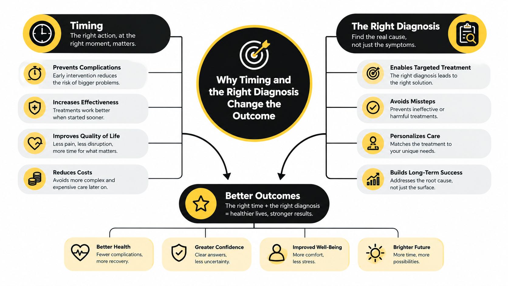
A diagnosis-specific decision tree is more useful than a fixed ladder. The examination, imaging, and nerve studies should point toward the next step. Sometimes that's rehabilitation. Sometimes it's a diagnostic block, decompression, or a coordinated evaluation of more than one compression site.
When to Seek Specialist Care and What to Expect Next
Arrange specialist evaluation when numbness keeps returning, pain follows an arm or leg pathway, symptoms interfere with sleep or work, or weakness is developing. Seek urgent medical attention for bowel or bladder changes, rapidly progressive weakness, major gait changes, or severe escalating numbness. Back pain accompanied by unexplained weight loss also deserves prompt medical assessment.
Bring these items to your first pain clinic visit:
- Prior imaging: MRI, CT, X-ray, or ultrasound reports and available images.
- Medication list: Include current medicines, previous trials, allergies, and side effects.
- Pain diary: Note location, triggers, sleep effects, weakness, and what relieves symptoms.
- Procedure history: List prior injections, blocks, therapy, surgery, and the response to each.
The consultation usually combines a focused neurological examination with imaging review and a discussion of options. A same-day steroid injection may involve brief observation afterward, while a stimulator pathway generally includes a staged trial before any permanent implant is considered. Recovery depends on the procedure and the underlying diagnosis, so your clinician should explain restrictions and milestones specifically rather than promise a universal timeline.
Insurance steps can also affect scheduling. This practical guide to prior authorization in clinics explains why documentation and payer review may be needed before some procedures. For patients in Chicago Ridge, Palos Heights, Palos Hills, Evergreen Park, and Hickory Hills, coordinating records among primary care clinicians, surgeons, rehabilitation providers, and a pain clinic helps keep the plan continuous.
Midwest Pain & Wellness provides diagnosis-focused, opioid-sparing care for spine and peripheral nerve conditions, including image-guided injections, nerve blocks, stimulation, radiofrequency ablation, and minimally invasive decompression when appropriate. Visit Midwest Pain & Wellness to request an evaluation and discuss which nerve compression treatment pathway fits your symptoms, examination, and imaging.