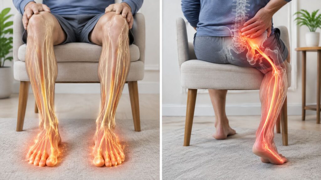
One review of sacral lateral branch radiofrequency neurotomy recorded 29 adverse events, including 3 serious events, among 1,367 patients. That figure describes one specific procedure and group of studies. It can't tell you the risk of heart, thyroid, knee-nerve, or other spine procedures. The review's findings show why a useful safety estimate needs to match the treatment being considered.
Radiofrequency ablation (RFA) uses heat to interrupt signals in a selected nerve or treat tissue in a specific organ or heart structure. Because the target and technique differ, radiofrequency ablation complications aren't captured by one universal rate. When you discuss risk, ask what procedure the number describes, how many people it includes, what researchers counted, and how long they followed patients.
Table of Contents
- Complication Rates by Type of Radiofrequency Ablation
- How to Read Complication Rates Fairly
- Rare Serious Complications and How They Happen
- Risk Factors That Change Your Odds
- How Care Teams Prevent Complications Step by Step
- Expected Effects, Warning Signs and Recovery
- Confirm Your Risk Before Booking RFA in Illinois
Complication Rates by Type of Radiofrequency Ablation
Reported complication rates differ by the nerve, organ, or heart structure treated. The table shows results from specific procedures and studies. Match the row to the treatment under consideration, then read the study context alongside the rate.
| Procedure | Reported complication rate | Most serious events | Study context |
|---|---|---|---|
| Atrial-fibrillation catheter ablation | Under 4% overall by 2015, compared with about 6% in earlier experience | Cardiac tamponade, stroke, and atrio-esophageal fistula; worldwide mortality was about 0.15% | Safety review |
| Thyroid nodule RFA | About 2%–3% postoperative complications; under 1% permanent or severe injury | Vocal-cord paralysis, hematoma, and nodule rupture | Reported overall rates ranged from 0% to 16.7% across studies |
| Sacral lateral branch neurotomy | 29 adverse events among 1,367 patients, including 3 serious events | Serious events were uncommon in the reviewed studies | Systematic review |
| Basivertebral nerve RFA | 15 treated participants, or 10.7%, had adverse events | Transient deficits and one retroperitoneal hemorrhage | Review |
| Genicular nerve RFA, 30-day window | Infection 0.1%; nerve injury 0%; deep-vein thrombosis (DVT) 2.0%; pulmonary embolism (PE) 1.2% | Thromboembolic events | National matched-cohort analysis |
| Cooled-RF neurotomy | One third-degree burn among 40 patients, or 2.5% | Skin burns | Systematic review |
| Bone-metastasis RFA with kyphoplasty or vertebroplasty | 16 of 583 patients, or 3%, had procedure-related adverse events | Increased pain or numbness affected about 1% | Review of 9 studies |
| All radiofrequency ablation, pooled | 10.9% | Not separated by procedure type | 2023 meta-analysis; microwave ablation was 27.4% |
The cardiac figures apply to catheter ablation for atrial fibrillation. A safety review reported overall complications under 4% by 2015, compared with about 6% in earlier experience, and worldwide mortality of about 0.15%. Serious events included cardiac tamponade, stroke, and atrio-esophageal fistula. These figures do not transfer to joint or spine RFA. The cardiac safety review
The noncardiac rows also show why procedure-specific context matters. For genicular-nerve RFA, a national matched-cohort analysis reported outcomes within a 30-day window. For bone-metastasis procedures, a review recorded procedure-related adverse events in 16 of 583 patients. The populations, targets, and study designs differ, so the percentages answer different questions. The genicular-nerve analysis and the bone-metastasis safety summary describe those separate settings.
The pooled 10.9% rate combines RFA procedures without separating them by treatment type. The same meta-analysis reported 27.4% for microwave ablation, a different technique. The 2023 comparison provides a broad reference point, while the row matching your procedure is more relevant to your decision. Check the number of patients behind the rate and the period researchers followed them.
How to Read Complication Rates Fairly
A complication percentage has meaning only when you know who was counted, what researchers called a complication, and how long they tracked patients. Those details matter because radiofrequency ablation can target different nerves, organs, or heart structures. A rate for one procedure is not automatically a useful estimate for another.
The denominator changes the apparent rate. In a cooled-radiofrequency review, one burn among 40 patients equaled 2.5%. If the same single event occurred among 1,367 patients, the rate would be under 0.1%. These examples show why the number of patients behind a percentage matters as much as the event count. The review of cooled-radiofrequency neurotomy reports the burn among 40 patients.
What the rate includes
Studies may count short-term soreness, numbness, or nerve irritation alongside lasting injury. When these outcomes are grouped together, the overall rate does not show how often permanent harm occurred. For example, a bone-metastasis review reported increased pain or numbness in about 1% of patients. That figure alone does not establish how long symptoms lasted or whether treatment was needed.
Follow-up time also shapes the result. A study tracking patients for 30 days may not capture symptoms that emerge or persist later, including nerve symptoms or changes in a thyroid nodule. Check whether the reported rate covers events during treatment, soon afterward, or across a longer follow-up period. This overview of radiofrequency ablation success rates explains how outcome measures affect interpretation.
Comparisons between technologies need the same care. A meta-analysis of cryoballoon and radiofrequency ablation for persistent atrial fibrillation found no statistically significant difference in total complications. Its odds ratio was 1.08, with a 95% confidence interval from 0.74 to 1.58. Since the interval includes 1.0, the observed difference could reflect chance; the result does not prove either approach is safer. The indexed comparison illustrates why a single point estimate cannot answer the safety question on its own.
Ask at your consultation: How many of this procedure does the team perform? How does the clinic record complications? What rate applies to the exact target being treated?
Rare Serious Complications and How They Happen
A serious complication depends in part on where the electrode is placed and what lies nearby. Radiofrequency ablation (RFA) sends current through a planned circuit, while a needle or catheter brings energy close to a target. If the planned circuit is compromised, current can pass through superficial tissue. A needle that strays from its intended path can also injure nearby structures.
When current or heat reaches the wrong tissue
A grounding pad that loses contact with the skin can interfere with the electrical circuit. Clinicians check the pad and electrode placement before delivering energy because poor contact can allow heat to affect tissue away from the target. A burn may require wound care and time to heal. As noted earlier, reports of burns after cooled-radiofrequency treatment describe a possible equipment or grounding problem, but the circumstances of those cases do not establish what will happen in an individual procedure.
Needle placement carries a different risk. In its review of basivertebral nerve RFA, The basivertebral nerve review describes a retroperitoneal hemorrhage associated with misdirected access through the pedicle, as well as temporary motor or sensory changes that resolved. The pedicle is part of the vertebra, and a route that passes too far from the planned path can reach tissues behind the abdominal cavity. Reviewing imaging and mapping the access route helps clinicians reduce the chance of this kind of injury. If a complication occurs, the response depends on the problem: bleeding may need assessment and treatment, while new weakness or numbness may prompt neurological evaluation.
Why the risk depends on the target
The structures around a knee nerve, a spinal target, and the heart are different, so their possible complications differ too. The genicular-nerve cohort and cooled-radiofrequency review discussed earlier describe events linked to those specific procedures and their study follow-up periods. Their findings should not be treated as rates for other targets.
Cardiac catheter ablation involves structures that are not near a knee or spinal treatment site. Serious concerns include cardiac tamponade, stroke, phrenic-nerve injury, and atrio-esophageal fistula, a connection between the heart and esophagus. The cardiac ablation review noted these events in its discussion of risk. Their relevance is specific to cardiac procedures, not a general measure of risk across all RFA.
Thyroid ablation has its own possible complications, including vocal-cord paralysis and nodule rupture. The practical question is which nearby structures matter for the planned treatment, what symptoms could signal a problem, and how the care team would assess them. Before consenting, ask the clinician to name the serious complications associated with your target and explain which symptoms need prompt attention.
Risk Factors That Change Your Odds
A patient taking a blood thinner who also has spinal hardware needs a different assessment from a patient without those factors, even if both are considering the same procedure. An average rate can't account for your medications, anatomy, or health history, so the treating team should review those details before scheduling.
| Risk factor | Why it may matter | What the team checks |
|---|---|---|
| Blood thinners or antiplatelet drugs | Bleeding or hematoma at the needle site | A written plan from the prescriber for holding and restarting medication |
| Osteoporosis | Vertebral or sacral fracture in spine procedures | Bone quality and anatomy on MRI or CT |
| Prior spine surgery or implanted hardware | Altered anatomy or scar tissue near the target | Imaging of implants and the proposed access route |
| Diabetes | May affect healing and infection risk | Glucose management and infection screening |
| Active infection near the site | May require delaying a procedure | Whether the infection has cleared |
| Pregnancy | Imaging decisions may need special consideration | Alternative timing or imaging plan |
| Peripheral neuropathy | Existing numbness can make new symptoms harder to distinguish | Baseline sensory testing before treatment |
These checks help the clinician decide whether to proceed, change the plan, or discuss another option. For vertebral or basivertebral nerve procedures, reviewing MRI or CT for pedicle anatomy, cortical integrity, nearby blood vessels, and neural structures is particularly relevant when osteoporosis, prior surgery, or altered anatomy is present. The aim is to understand the access route and your starting point before treatment.
Don't stop a blood thinner on your own. Ask the prescribing clinician and the treating team to agree on a written hold-and-restart plan. Bring a complete medication list, including antiplatelet drugs and supplements, so the team can review your full regimen.
Neuropathy deserves a direct conversation. If you already have numbness, tell the clinician where it occurs and whether it has changed; baseline sensory testing gives the team a clearer reference for checking you afterward.
Bring your imaging reports, operative notes, current medication list, and records of any prior nerve or spine procedures to the consultation. Those details let the team discuss risks for your anatomy and treatment target, instead of relying only on a general rate.
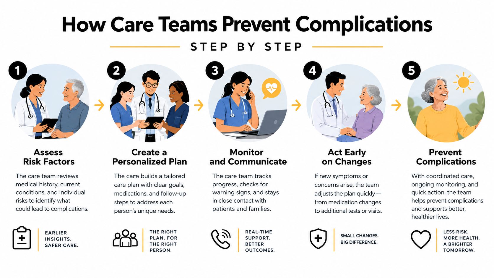
How Care Teams Prevent Complications Step by Step
Eight checks run from confirming the treatment target to planning aftercare. Each addresses a different part of the procedure, such as needle placement, electrical current, or response to a new symptom. Their purpose is to reduce avoidable risks before energy is delivered and to make changes easier to recognize during and after treatment.
Review MRI or CT. The clinician checks pedicle anatomy, bone integrity, nearby blood vessels, and nerves, especially when bone is fragile or prior surgery has changed the anatomy. This review helps identify a needle path that avoids vulnerable structures.
Reconcile medications and health concerns. The team documents a hold-and-restart plan for blood thinners or antiplatelet drugs, reviews glucose when relevant, and checks for signs of infection. If a concern needs attention, the clinician can address it before proceeding.
Confirm the target and baseline symptoms. The team verifies the intended level and records sensory and motor findings before treatment. These baseline observations provide a point of comparison if symptoms change afterward.
Check the grounding pad and electrode. Staff confirm that the pad has full skin contact and that the electrode is secure before energy is delivered. Good contact helps keep the current on its intended path.
Place the cannula with imaging guidance. The clinician uses imaging to guide the needle and avoid non-target motor roots and major vessels. The planned path should fit the patient's anatomy.
Test stimulation when appropriate. Low-intensity stimulation can help confirm the target nerve and check for an unintended motor response before lesioning. If the response differs from what the clinician expects, needle position can be reassessed.
Monitor comfort and vital signs. The team observes the patient during and after treatment and records any new deficit. Monitoring gives staff a chance to respond if a concerning change occurs.
Give written aftercare instructions. Before leaving, the patient should receive warning signs, a same-day contact number, and guidance on when emergency care is needed. Written instructions help clarify whom to contact and how quickly to act.
Patients can ask how the team will confirm the target, record baseline symptoms, check pad contact and needle position, and monitor them during treatment. Ask for the contact number to use afterward, too. For details about radiofrequency ablation for spine pain, discuss the planned approach with the clinician familiar with your diagnosis.
Expected Effects, Warning Signs and Recovery
After RFA, patients typically have soreness near the needle sites, bruising, temporary numbness, or a localized burning feeling similar to sunburn. With lumbar facet or medial-branch neurotomy, burning around the treated sensory nerve is often post-procedure neuritis, or nerve irritation. It generally resolves within 2–4 weeks, according to a clinical reference on lumbar radiofrequency neurotomy. New or persistent radiating symptoms need assessment.
Bruising should settle over time. A bruise that spreads or becomes more painful after the first week needs a call to the treating clinic. The same applies to numbness or nerve discomfort that worsens instead of easing.
Recovery depends on the procedure
Recovery varies with the target being treated. The table describes typical patterns and warning signs by procedure. Use it to understand what to watch for, then follow your clinician's guidance for your treatment.
| Procedure | Typical soreness | Relief window | Watch for |
|---|---|---|---|
| Facet or medial-branch neurotomy | Burning or sunburn-like soreness that may last 2–4 weeks | Relief may build over several weeks | Escalating weakness or progressive numbness |
| Genicular nerve RFA | Soreness around the treated area | Relief may build gradually | One-sided calf swelling, chest pain, or breathlessness |
| Basivertebral nerve RFA | Incision or back pain | Relief may take time to develop | Trouble urinating or new leg weakness |
| Thyroid nodule RFA | Neck tenderness | The nodule may shrink gradually | Hoarseness that persists or worsens |
| Cardiac AF catheter ablation | Chest discomfort | Return to activity depends on the care team's guidance | Chest pain, shortness of breath, or stroke signs |
For more detail on the discomfort patients may experience during and after treatment, read this guide to whether radiofrequency ablation hurts. Activity and medication advice varies by treatment target and personal health factors, so follow the instructions from your own clinician.
Know when to call
Call the treating clinic the same day for fever, drainage, or spreading redness at the treatment site; worsening weakness or numbness; one-sided calf swelling; new hoarseness after thyroid treatment; or trouble urinating after a spine procedure. Contact the clinic promptly if symptoms are getting worse rather than waiting for a routine follow-up.
Call 911 for an emergency such as chest pain, sudden shortness of breath, sudden weakness, facial droop, or other stroke signs after cardiac ablation. These symptoms require immediate assessment.
Treatment depends on the complication. Neuritis may be managed with conservative care, anti-inflammatory medication, and reassessment. Persistent radiating symptoms may call for imaging to check for nerve injury. A hematoma may need pressure and monitoring; a burn may require wound care over an extended period. Infection needs prompt medical assessment and may require antibiotics or drainage, while cardiac tamponade requires urgent drainage. If meaningful relief has not appeared by about three months, ask your clinician to review whether the diagnosis and treatment still fit your symptoms.
Confirm Your Risk Before Booking RFA in Illinois
RFA complication rates vary by the nerve, organ, or heart structure treated, so one headline number may not describe your procedure. Before deciding, ask whether the expected benefit fits your diagnosis and how RFA compares with medication, injections, or other options. Your health history and treatment target also shape the decision.
Use your consultation to clarify choices that are specific to you:
- Review medication and imaging plans. Ask what information the clinician needs about your medications and medical history, and whether any additional records or imaging are needed before treatment.
- Discuss the clinician's experience. Ask how often the physician performs the particular procedure you are considering and who will answer questions about your care.
- Clarify follow-up arrangements. Find out when and how the team will check on your recovery, whom to contact with a concern, and what to do if you cannot reach the clinic.
- Request written instructions. Leave with a clear record of your next steps, appointment details, and the person responsible for coordinating your care.
Midwest Pain & Wellness is a pain management clinic in Chicago Ridge, Illinois. It serves patients from Chicago Ridge and nearby communities, including Palos Heights, Palos Hills, Evergreen Park, and Hickory Hills. The clinic offers radiofrequency ablation and coordinates care with surgeons, primary care providers, and other clinicians.
Before scheduling, request a written, procedure-specific risk review that explains how your treatment plan fits your target and health history. A consultation gives you time to raise concerns and decide whether RFA is appropriate for your situation.
Midwest Pain & Wellness offers pain management services, including radiofrequency ablation, for people seeking an individualized discussion of treatment options and procedure-specific risks. If you're in Chicago Ridge, Palos Heights, Palos Hills, Evergreen Park, or Hickory Hills, visit Midwest Pain & Wellness to request a consultation before scheduling.