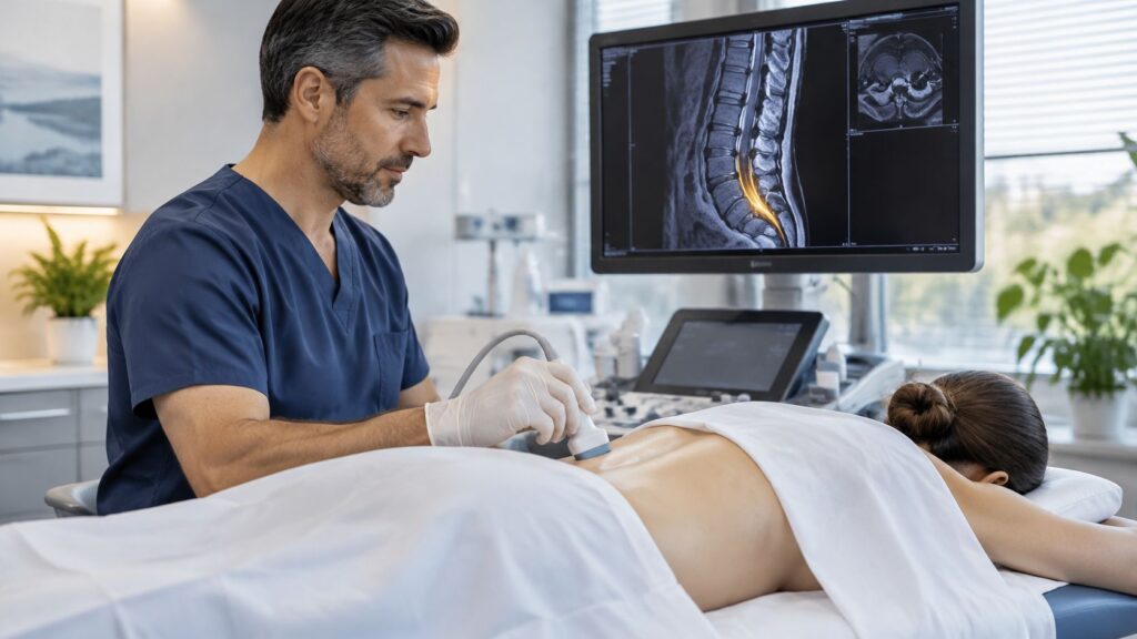
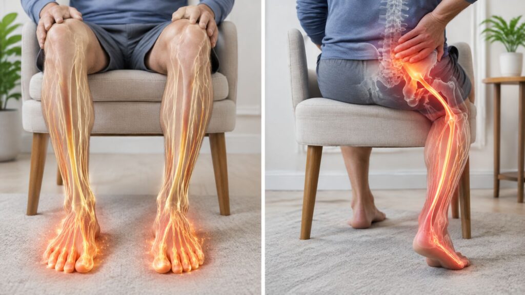
Sacroiliac joint dysfunction is pain generated by the joint where the spine meets the pelvis, and research suggests it may account for roughly 15% to 30% of chronic low back pain cases. It's often missed because its symptoms can resemble disc, facet joint, or hip pain.
You may be dealing with that familiar pattern right now: pain just below the beltline, worse when you stand from a chair, roll in bed, climb stairs, or shift your weight onto one leg. Perhaps an MRI showed a disc bulge, treatment focused on the lumbar spine, and yet your pain never received a convincing explanation.
The sacroiliac joint, or SI joint, can be the source, but I don't treat the phrase “SI joint dysfunction” as a complete diagnosis by itself. The pain may come from the joint surface, the ligaments around it, or the muscles that help control pelvic movement. That broader SI joint complex explains why symptoms can persist even when a scan looks ordinary.
This distinction matters in Illinois communities such as Chicago Ridge, Palos Heights, Palos Hills, Evergreen Park, and Hickory Hills, where patients often arrive after months of treatment aimed at the wrong structure. The right approach is to identify the pain pattern, examine competing causes, confirm the suspected source when appropriate, and then escalate treatment in a measured order.
Table of Contents
- Understanding Sacroiliac Joint Dysfunction
- How the SI Joint Works and Why It Hurts
- Common Causes and Who Is Most at Risk
- Recognizing SI Joint Pain Versus Other Back Pain
- How Clinicians Diagnose SI Joint Dysfunction
- Evidence-Based Treatment Options From Conservative to Interventional
- Prognosis, Self-Care, and When to See a Specialist
Understanding Sacroiliac Joint Dysfunction
A 42-year-old runner comes to clinic with three years of “lower back pain.” Previous clinicians blamed a disc. Lumbar imaging looks unremarkable, and physical therapy designed for a herniated disc never changes the pain. The discomfort sits on one side near the back of the pelvis, flares during single-leg loading, and makes turning in bed surprisingly difficult.
That story is common because the SI joint sits at a crossroads. It connects the sacrum, the triangular bone at the base of the spine, to the ilium, the large pelvic bone. Each person has an SI joint on either side. When pain arises from this region, patients may describe it as low back, buttock, hip, or upper-thigh pain rather than identifying the joint itself.
The SI joint complex
Sacroiliac joint dysfunction means the SI region is producing pain because of irritation, altered movement, inflammation, instability, or stress on nearby structures. The useful clinical concept is broader than the joint cavity alone. It includes:
- The joint surfaces, which can become irritated or arthritic.
- Supporting ligaments, which can become strained or lax.
- Surrounding muscles, including the gluteal and deep hip muscles that help transfer load.
- Nearby pain-sensitive tissues, which can refer discomfort into the buttock, groin, or thigh.
This is why a normal MRI doesn't automatically rule out SI joint complex pain. A scan may help exclude other problems, but it often can't prove that the SI region is innocent or guilty.
Why clinicians miss it
Low back pain itself has a reported lifetime prevalence of 85%, according to an NIH-hosted review of low back pain epidemiology. Within that large group, the SI joint may account for about 15% to 25% of axial low back pain patients, while other reviews estimate SI-related pain at 15% to 30% of low-back-pain cases, as summarized in the same clinical literature.
Those ranges aren't contradictory. They reflect different patient populations and diagnostic methods. The practical point is that a meaningful portion of people with persistent, nonradicular, or one-sided buttock pain may have SI joint dysfunction rather than primary disc or facet disease.
Practical rule: If treatment aimed at the lumbar disc repeatedly fails, the diagnosis deserves to be reopened, not simply intensified.
How the SI Joint Works and Why It Hurts
Think of the SI joint as the keystone of a bridge. The spine sits above it, the pelvis and legs support it below, and the joint transfers force between those structures whenever you walk, run, stand, or lift.
The sacrum acts like a wedge between the two pelvic bones. Strong ligaments hold it in place, while the gluteals, piriformis, abdominal muscles, and thoracolumbar fascia help control movement around the pelvis. The joint isn't designed for large motion. Its limited movement is important because it allows the pelvis to absorb and redirect force without becoming unstable.
Small movements with large consequences
The SI joint performs subtle motions often described as nutation and counternutation. During nutation, the upper sacrum moves in a way that increases pelvic stability. Counternutation reverses that relationship. These movements are small, but they influence how forces travel from the spine into each leg.
Pain may develop when the joint complex becomes either too mobile or too restricted:
- Hypermobility means the ligaments and surrounding tissues don't control motion effectively. Pregnancy and postpartum pelvic changes can contribute to ligamentous laxity.
- Hypomobility means the joint and nearby tissues become stiff. Degenerative change, arthritis, or protective muscle guarding can limit normal movement.
- Inflammation can make ordinary loading painful even when the amount of movement hasn't changed dramatically.
- Muscle imbalance can shift more force onto one side of the pelvis, particularly during running, lifting, or prolonged standing.
The body then compensates. A patient may shorten a stride, rotate the trunk, lean away from the painful side, or overuse the hip and lumbar muscles. Those adaptations can create secondary pain, which makes the original source harder to identify.
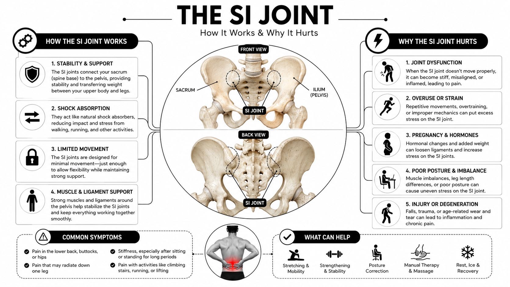
The most useful mental model is not “a joint that is out of place.” It's a load-transfer system that has become irritated, poorly controlled, inflamed, or mechanically overloaded. That framing also explains why strengthening, movement retraining, injections, and nerve-targeted procedures serve different purposes.
Common Causes and Who Is Most at Risk
SI joint dysfunction usually develops from a combination of loading, tissue sensitivity, and altered mechanics. A fall or motor vehicle accident can strain the ligaments directly. Repetitive activities, including running or heavy lifting, can repeatedly load one side of the pelvis, especially when technique, strength, or recovery is inadequate.
Pregnancy deserves special attention because the pelvis must accommodate changing load and ligamentous laxity. Some patients improve after delivery, while others continue to experience pain because the muscles and movement patterns never fully regain control. The American Academy of Family Physicians review linked SI joint dysfunction with greater SI joint mobility and load-related ligament strain in women, and noted that women present more often than men in clinical settings. The same review is discussed in the clinical evidence summarized through PubMed.
Inflammatory arthritis, including ankylosing spondylitis, can affect the SI joints differently from mechanical dysfunction. Morning stiffness, persistent inflammatory symptoms, and pain unrelated to ordinary loading require a broader medical evaluation. Osteoarthritis may also make the region stiff and painful, particularly as tissues lose their ability to distribute force smoothly.
Why prior spine surgery matters
Lumbar fusion changes how the spine and pelvis share motion and load. The SI joints may then receive more stress, particularly after multi-level fusion. That doesn't mean every patient with post-fusion pain has SI dysfunction. It means the SI region should be assessed rather than assuming the surgical level remains the only possible source.
A leg length discrepancy, hip problem, or altered gait can create asymmetric pelvic loading. Athletes in sports that repeatedly favor one side, such as cutting, kicking, or rotational activities, can develop similar imbalances. Pelvic or sacral trauma adds another direct source of ligament and joint irritation.
| Risk Factor | Mechanism | Most Affected Population |
|---|---|---|
| Trauma | Ligament strain, joint irritation, or altered force transfer | Patients after falls, collisions, or pelvic injury |
| Repetitive loading | Repeated asymmetric stress during impact or lifting | Runners, field athletes, and heavy-lifting workers |
| Pregnancy and postpartum change | Increased pelvic mobility and changing load distribution | Pregnant and postpartum patients |
| Inflammatory arthritis | Inflammation involving the SI region | Patients with inflammatory rheumatic disease |
| Prior lumbar fusion | Greater stress transferred to the pelvic ring | Patients after lumbar fusion, especially multi-level procedures |
| Leg length or gait asymmetry | Uneven loading across the pelvis | Patients with limb, hip, knee, or gait problems |
| Degenerative change | Stiffness, surface irritation, and reduced load tolerance | Older adults with osteoarthritic changes |
Women aged 30 to 60, postpartum patients within the first year, post-lumbar-fusion patients, athletes in asymmetric sports, and people with prior pelvic or sacral trauma are commonly represented in clinic populations. These are risk patterns, not diagnoses. The examination still has to connect the history to the pain behavior.
Recognizing SI Joint Pain Versus Other Back Pain
SI joint pain often has a recognizable location. Patients commonly point to an area just off the midline over the posterior superior iliac spine, often within 3 centimeters of that bony landmark. The pain may spread into the buttock, posterior thigh, groin, or upper thigh, but it generally doesn't behave like nerve-root pain traveling below the knee.
Transitions are especially revealing. Standing from a chair, rolling in bed, climbing stairs, walking uphill, bending, twisting, or shifting weight onto one leg can reproduce the familiar pain. Prolonged sitting or standing may also aggravate it. These clues increase suspicion, but no single symptom proves the SI joint is responsible.
A practical pattern comparison
| Feature | SI Joint | Disc Herniation | Facet Joint | Hip Osteoarthritis |
|---|---|---|---|---|
| Main location | One-sided pain below the beltline, near the posterior pelvis | Low back pain with possible leg-dominant symptoms | Central or one-sided lumbar pain | Deep groin pain, sometimes extending to the thigh |
| Referral pattern | Buttock, posterior thigh, groin, or upper thigh | Often follows a nerve-root distribution | Usually local or near the buttock | Groin, front of thigh, or knee |
| Numbness or weakness | Uncommon | More concerning when it follows a nerve pattern | Uncommon | Usually limited by pain rather than nerve deficit |
| Common aggravators | Sit-to-stand, stairs, rolling in bed, single-leg loading | Sitting, bending forward, coughing, or straining may worsen symptoms | Extension, standing, or rotation may provoke pain | Walking, pivoting, and hip rotation |
| Key patient clue | Pain is off-center and transition-related | Leg symptoms may dominate the complaint | Back pain worsens with extension | Groin-deep pain and reduced hip motion |
True radicular symptoms deserve careful attention. Dermatomal numbness, progressive weakness, or clear nerve-pattern pain can point toward lumbar nerve compression rather than isolated SI joint dysfunction. Hip osteoarthritis more often causes deep groin pain and painful restriction of hip movement, although hip and SI disease can coexist.
Facet pain can also overlap, particularly when pain increases with extension and rotation. A patient may have more than one pain generator, which is why self-screening is useful for preparing a clinical history but isn't enough for choosing an injection or procedure.
The most helpful description isn't “my back hurts.” Tell the clinician where the pain starts, what movement reproduces it, whether it travels below the knee, and whether numbness or weakness accompanies it.
How Clinicians Diagnose SI Joint Dysfunction
A patient may point to one side of the low back or buttock and report pain after a fall, pregnancy, surgery, or a change in activity. The first task is to determine whether the complaint fits the SI joint complex, which includes the joint, supporting ligaments, and nearby muscles, or whether the dominant source is the lumbar spine, hip, or a nerve.
I start with a targeted history. The location, onset, aggravating movements, and load-transfer activities matter. I also ask about numbness, weakness, and pain below the knee, because those findings can shift the evaluation toward nerve or lumbar disease.
The examination uses a group of provocative maneuvers, rather than one supposedly decisive test. Common tests include FABER, thigh thrust, Gaenslen, compression, and distraction. FABER places the hip in flexion, abduction, and external rotation. Thigh thrust loads the SI region through the femur. Gaenslen stresses the pelvic ring, while compression and distraction change forces across the joint complex and its supporting tissues.
A commonly cited rule is that three or more positive provocation tests out of five raises suspicion, as described in the American Academy of Family Physicians review of SI joint dysfunction. It does not confirm that the SI joint is the only pain generator. These maneuvers are more useful for lowering suspicion when they are consistently negative than for proving a single diagnosis.
What imaging can and cannot do
Imaging helps identify competing causes and structural problems, but a scan cannot reproduce the patient's familiar pain during ordinary movement.
- X-rays assess alignment, degenerative change, and some structural abnormalities.
- MRI can show inflammatory sacroiliitis, marrow edema, disc disease, or other pathology.
- CT clarifies bony anatomy and may help when fusion hardware limits other views.
Clinical history, examination, and imaging may still leave uncertainty. For that reason, small-volume anesthetic blocks became an important diagnostic method in the detailed review of SI joint pain in Anesthesia & Analgesia.
The confirmatory step
An image-guided diagnostic SI joint injection places local anesthetic into the suspected joint under fluoroscopic guidance. The practical question is whether numbing that structure temporarily reduces the patient's typical pain during movements that normally provoke it. Information about a fluoroscopy-guided injection explains how imaging supports procedural accuracy.
If relief is borderline, the clinician may repeat the block on another day. The diagnosis should combine the history, examination, exclusion of competing causes, and response to a properly performed block. An MRI finding alone is not enough.
Patients comparing approaches to persistent back pain can browse chronic back pain solutions, but an SI-specific concern still requires a focused evaluation.
Evidence-Based Treatment Options From Conservative to Interventional
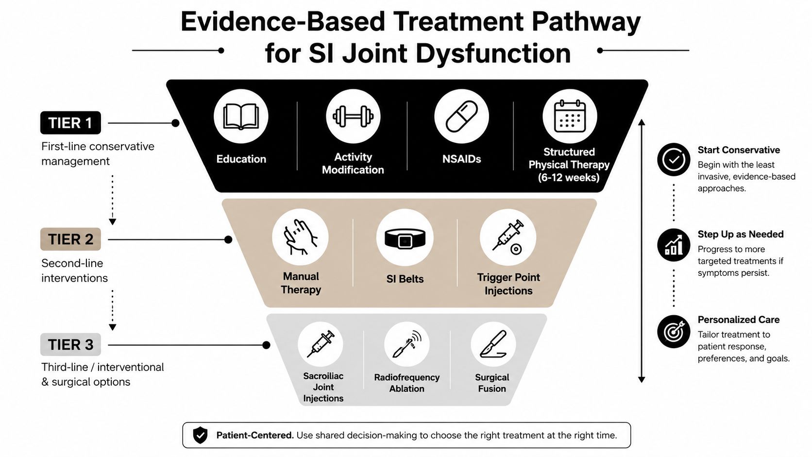
Treatment should follow an escalation pathway, not a menu in which every patient receives the same procedure. The first question is whether the pain generator has been reasonably identified and whether the patient can participate in rehabilitation.
Tier one focuses on control and capacity
Education, activity modification, anti-inflammatory medication when medically appropriate, and structured physical therapy form the foundation. A targeted program should address pelvic stabilization, trunk control, gluteal strength, and hip external rotator function. The purpose isn't to force the SI joint into a particular position. It's to improve how the surrounding system handles load.
A patient may need to temporarily reduce running hills, heavy asymmetric lifting, prolonged sitting, or repeated twisting. That isn't permanent avoidance. It's a way to reduce aggravation while strength and movement tolerance improve.
Tier two adds targeted treatment
Manual therapy, an SI belt, and trigger-point treatment may help selected patients, particularly when muscle guarding or external instability contributes to symptoms. An image-guided intra-articular corticosteroid injection can serve two roles. It may reduce inflammation and provide therapeutic relief, but it also helps test whether the joint is involved.
I explain the limitation plainly: an injection may help for weeks or months, but it doesn't correct every mechanical or muscular contributor. Recent reviews conclude that intra-articular injections are generally more useful diagnostically than therapeutically, as summarized in the systematic review of SI joint interventions.
Tier three and beyond
For patients with confirmed SI-region pain who obtain temporary benefit from blocks but continue to have significant symptoms, cooled or conventional radiofrequency ablation may be considered. These procedures target lateral branch nerves that carry pain signals from the SI joint complex. Recent reviews describe cooled radiofrequency as appearing particularly effective for pain and function, while also emphasizing that high-quality evidence remains limited.
Platelet-rich plasma and other regenerative approaches have limited but growing evidence. They shouldn't be presented as guaranteed cartilage repair or a substitute for diagnosis.
Minimally invasive SI joint fusion belongs at the end of the pathway. It may be reasonable for carefully selected patients with a confirmed source, persistent functional limitation, and failure of appropriate nonoperative and interventional care. The available trial literature, including iFuse and SImmetry outcomes, supports discussion in selected cases, not automatic surgery for every patient with SI pain.
For broader perspectives on holistic chronic pain relief, patients may find value in considering sleep, stress, conditioning, and overall health alongside targeted procedures. If an injection is being discussed, understanding what a nerve block injection is can make the consent conversation more productive.
Prognosis, Self-Care, and When to See a Specialist
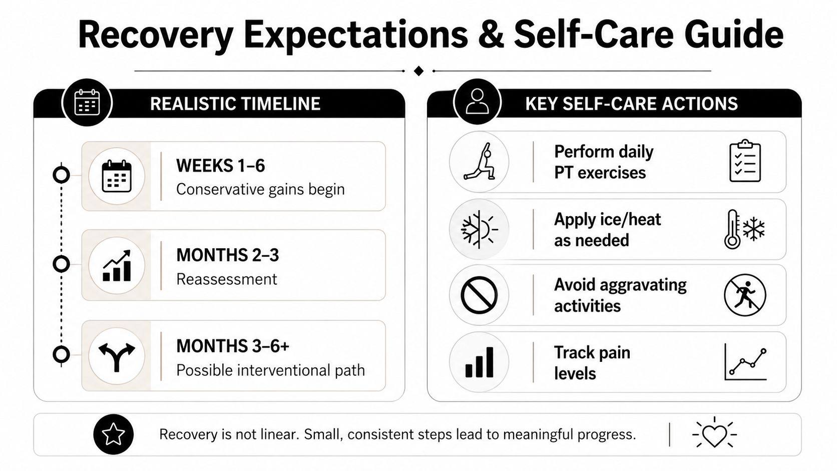
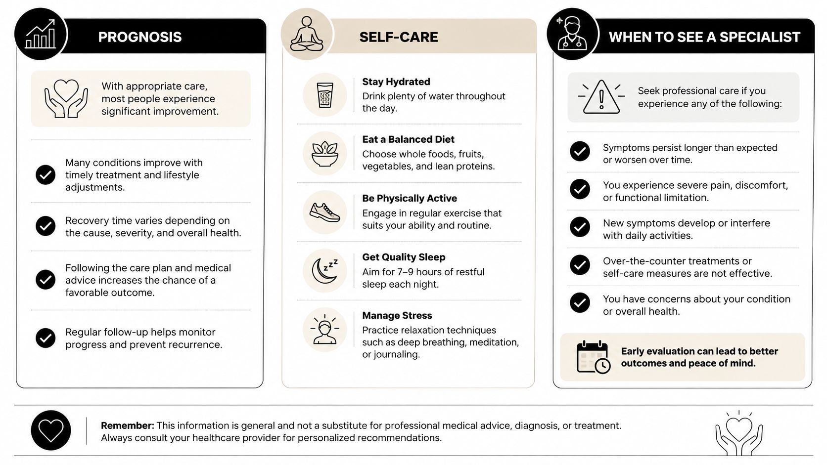
Most patients improve through a layered plan, but recovery is rarely immediate. Conservative gains usually build over weeks, while an interventional treatment may require follow-up over months to determine whether it changed function as well as pain.
Prognosis becomes less favorable when smoking, untreated hip or lumbar disease, ongoing ligamentous laxity, poor conditioning, or repeated aggravating activity continues to stress the SI complex. Those factors don't make recovery impossible. They tell us that an injection alone is unlikely to be a complete solution.
What you can do during a flare
- Use relative rest: Reduce the movement that triggers pain without staying completely inactive.
- Choose heat or ice: Use whichever provides better relief. Ice may feel useful after an acute aggravation, while heat can relax guarded muscles.
- Change sleep position: Side sleepers often tolerate a pillow between the knees, while others do better with support under the knees when lying on the back.
- Practice pelvic control: Perform the stabilization exercises prescribed for your specific deficits rather than copying an aggressive online routine.
- Pace activity: Break lifting, walking, and household tasks into manageable intervals before pain escalates.
- Support general health: Weight management, sleep, and gradual conditioning can reduce repeated stress on the pelvis.
Seek medical attention promptly for saddle anesthesia, progressive weakness, fever, unexplained weight loss, bowel or bladder changes, significant night pain, or pain that persists beyond six weeks despite reasonable home care. These findings may indicate a problem that self-care cannot safely address.
Before a visit, write down where the pain starts, what makes it worse, whether it travels below the knee, and whether numbness or weakness occurs. The guide on how to describe pain to your doctor can help you organize that information.
For patients in Chicago Ridge, Palos Heights, Palos Hills, Evergreen Park, Hickory Hills, and nearby Illinois communities, the next step is a focused pain consultation. Midwest Pain & Wellness can evaluate competing spine, hip, and SI joint causes and discuss a diagnostic SI injection pathway when the examination supports it. Bring prior imaging, a medication and treatment history, and a short record of the movements that reproduce your pain.
If your low back or buttock pain keeps returning despite routine treatment, visit Midwest Pain & Wellness to request an evaluation with a double board-certified interventional pain specialist. The clinic offers personalized, opioid-sparing care, including image-guided SI joint injections and other targeted options when the diagnosis is confirmed.