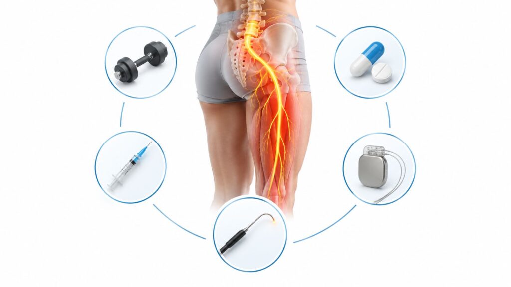
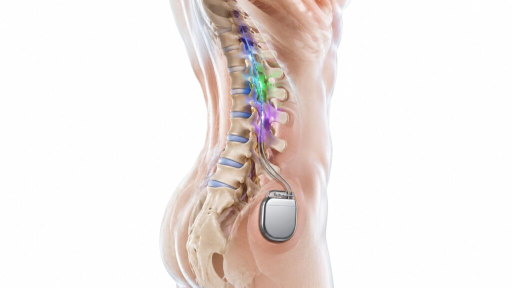
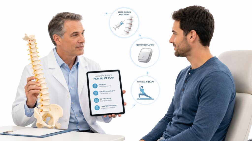
You feel the pain just to one side of your lower back, near the dimple above the buttock. Sitting through a meeting, standing from a chair, climbing stairs, or turning in bed makes it sharper, and the ache may travel into your hip or thigh. Because the pattern resembles sciatica or a lumbar strain, you may have already focused on the spine while the sacroiliac joint remained unexamined.
Sacroiliac joint pain causes aren't limited to one injury or one type of joint damage. Trauma, repetitive loading, pregnancy, inflammatory disease, degeneration, prior lumbar fusion, and irritation in the ligaments or muscles around the joint can all produce similar symptoms. A review estimates that SI joint pain accounts for about 15% to 30% of chronic, nonradicular low back pain, while another clinical review places SI joint dysfunction at approximately 25% in adults with chronic low back pain (clinical review of SI joint dysfunction).
Table of Contents
- When Low Back Pain Is More Than a Spine Problem
- How Your Sacroiliac Joint Works and Why It Hurts
- Trauma Repetitive Stress and Post Fusion Overload
- Pregnancy Degeneration and Inflammatory Causes
- Inside the Joint vs Around the Joint Pain Generators
- How SI Joint Pain Feels and How It Is Differentiated
- Next Steps for Lasting Relief in the Chicago Ridge Area
When Low Back Pain Is More Than a Spine Problem
A patient may report a deep ache below the beltline, usually more pronounced on one side. Sitting for a long time, standing on one leg, walking uphill, climbing stairs, or rising from a chair can intensify it. Pain may extend into the back of the thigh, creating concern about a pinched nerve.
That concern makes sense. The sacroiliac joint lies between the lower spine and pelvis, while nearby nerves, muscles, ligaments, and the hip can create overlapping symptoms. A sharp twinge during movement may become a dull ache afterward. Weight shifting from one leg to the other can also provoke a flare.
A useful distinction: Pain near the SI joint identifies an area to examine, not automatically the structure causing the pain.
Sacroiliac joint pain causes form a syndrome with several possible generators. Pain may arise from structures inside the joint, such as its surfaces or capsule, or from tissues around it, including ligaments and muscles. These sources can feel similar, yet each points to a different diagnostic path. Normal imaging does not rule out significant pain because scans may show bone and alignment more clearly than irritated soft tissues, altered movement, or pain sensitivity.
The SI joint is a major but often underrecognized source of chronic low back pain. Pregnancy, older age, inflammatory arthritis, previous spine surgery, trauma, and leg length discrepancy can all increase suspicion (review of prevalence and risk factors). For Illinois patients, that history helps a pain specialist decide whether to examine the joint itself, the surrounding tissues, the lumbar spine, or several areas together.
Until you are assessed, avoid repeatedly provoking the movement rather than pushing through a flare. Guidance on ways to avoid aggravating your spine may help you adjust daily activity while staying gently active.
Midwest Pain & Wellness is a pain and wellness clinic in Chicago Ridge, Illinois, serving Chicago Ridge, Palos Heights, Palos Hills, Evergreen Park, and Hickory Hills. A clear history of triggers, prior procedures, and pain location can make the next evaluation more focused and useful.
How Your Sacroiliac Joint Works and Why It Hurts
You have two sacroiliac joints, one on each side. Each connects the sacrum, the triangular bone at the base of the spine, with the ilium, the broad wing of the pelvis. Strong ligaments hold the bones together, and the joint is designed mainly for stability and weight transfer rather than the large range of motion found in the hip.
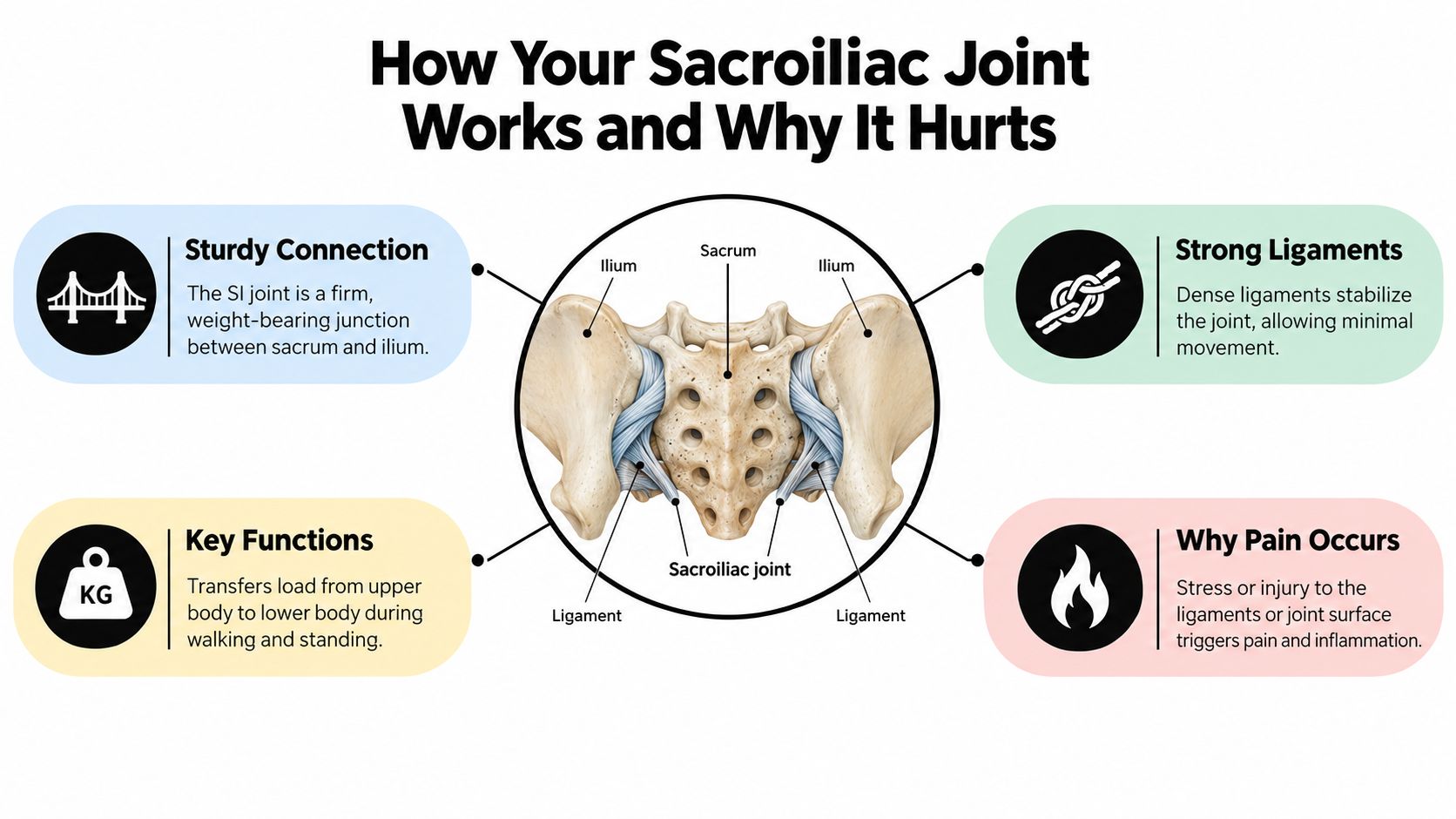
Think of the SI joint as a shock-absorbing bridge between your spine and legs. It doesn't swing freely like a hinge. Instead, it allows small, controlled movements while distributing forces each time you walk, stand, bend, or lift.
The load-transfer problem
Pain can begin when the joint moves too little, moves too much, or receives force unevenly. A stiff joint may pass stress to nearby ligaments and muscles. An overly mobile joint may strain the tissues that normally stabilize it. Shear and torsional forces can irritate the joint surface, the supporting ligaments, or both.
This is why an everyday movement can hurt even when you can't identify one dramatic injury. A change in gait, uneven leg length, altered pelvic mechanics, or increased loading after spine surgery can shift force toward one side of the pelvis. The body keeps moving, but the tissues may no longer share the work efficiently.
Why normal imaging doesn't settle the question
X-rays and other scans can show bone changes, fractures, advanced arthritis, infection, or other structural conditions. They may not reveal a painful ligament, a small soft-tissue injury, myofascial irritation, or a movement-related problem. A normal image can therefore be reassuring in one sense while still failing to explain severe pain.
A thorough review describes ligamentous injury, fractures, and myofascial pain as more common extra-articular sources than intra-articular causes. It also explains that hypo- or hypermobility, shear forces, micro- or macro-fractures, and adjacent-segment disease can stress the SI region (review of SI joint pain mechanisms).
Trauma Repetitive Stress and Post Fusion Overload
A fall onto one side, a vehicle collision, or one heavy lift can load the pelvis beyond what the SI joint and its supporting tissues can absorb. The injury may involve the joint surface, nearby ligaments, or both. Sometimes the event is obvious. Sometimes a seemingly minor incident is followed by pain that feels far greater than the original impact.
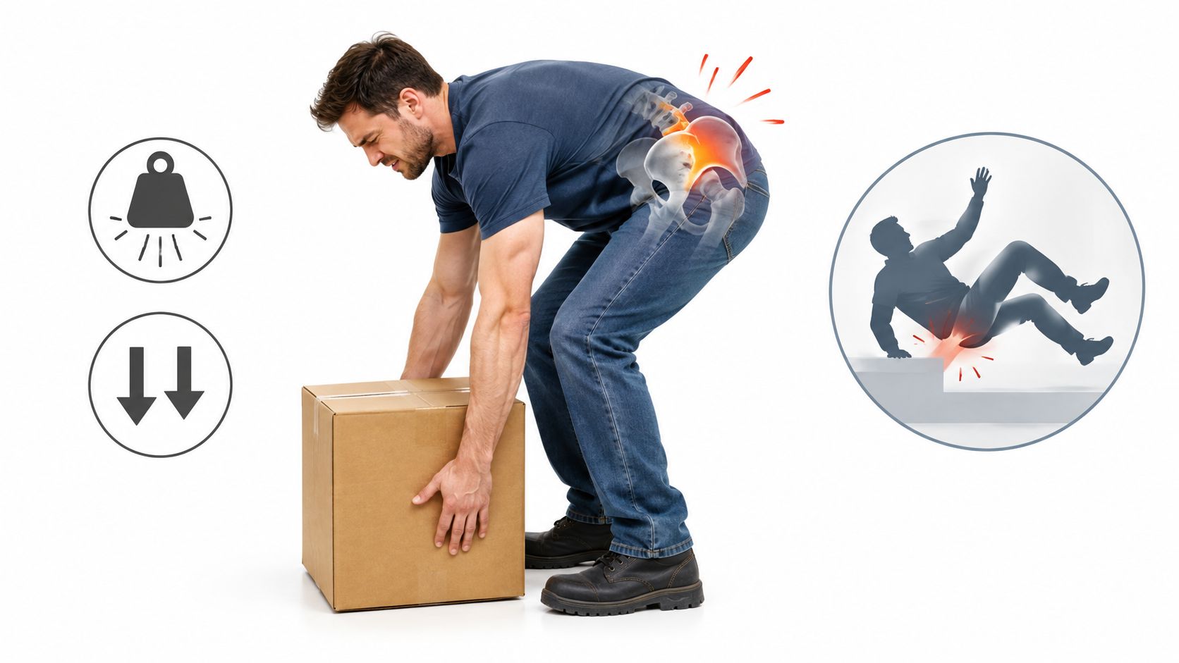
One retrospective study of 54 patients with injection-confirmed SI joint pain found trauma in 44% of cases, cumulative repetitive stress in 21%, and no specific identified cause in 35% (review summarizing traumatic and degenerative causes). Those figures describe one patient group, not every person with SI joint pain. They do explain why an evaluation covers both major injuries and repeated loading.
Repetitive stress can build quietly
Walking, standing, lifting, and twisting transfer force through the pelvis. If one side repeatedly carries more of that load, the stabilizing tissues may become irritated over time. Leg length discrepancy, obesity, hypermobility, a lasting gait change, and an old injury can all alter that distribution.
The onset may feel gradual rather than dramatic. Symptoms can appear after weeks or months of changed movement, increased activity, or compensation for a painful hip, knee, or lumbar spine. Intra-articular pain comes from structures within the joint, while extra-articular pain comes from ligaments and other tissues around it. That distinction matters because the painful area may need a different examination and treatment approach, even when both patterns feel like buttock or low back pain.
Pregnancy can also change pelvic loading and ligament stability. If you are researching baby brain development tips, keep the physical changes of pregnancy in view too, especially when pelvic or buttock pain starts during pregnancy or continues afterward.
Why lumbar fusion can shift stress to the SI region
A lumbosacral fusion stabilizes selected spinal segments, but it also changes how movement and force pass through the lower back and pelvis. The SI joint may then absorb more of the load that previously moved through the fused levels. Reviews report SI-joint prevalence of 32% to 37% after lumbosacral fusion surgery (clinical overview of SI joint dysfunction).
Persistent pain after surgery does not identify one cause by itself. The spine, SI joint, adjacent segments, nerves, and surrounding soft tissues may each require assessment. For Illinois patients, that referral question helps determine whether symptoms point toward the fused region, the SI joint, or an extra-articular generator.
Pregnancy Degeneration and Inflammatory Causes
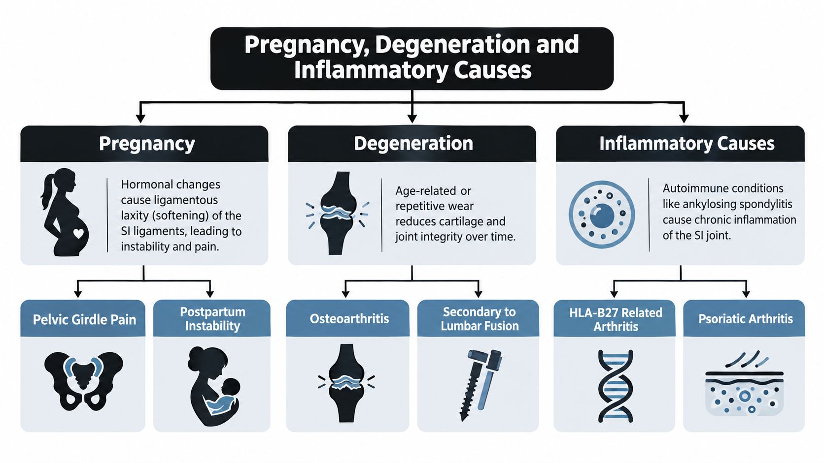
Pregnancy can shift SI joint loading before pain becomes obvious. Hormonal changes loosen supporting ligaments, while the growing body and altered gait change how force travels through the pelvis. The result may be irritation inside the joint, strain in the surrounding ligaments, or both. Symptoms can begin during pregnancy and continue after delivery.
Pregnancy is one recognized risk factor among several. Sacroiliac joint pain accounts for about 15% to 30% of chronic, nonradicular low back pain, with risk also associated with older age, inflammatory arthritis, previous spine surgery, trauma, and leg length discrepancy. This range describes a clinical syndrome, not one single disease, so the referral question remains important for Illinois patients. Pain that began with pregnancy may point toward pelvic loading and ligament stress, while symptoms with inflammatory features may require a broader medical evaluation. (review of SI joint prevalence and risk factors)
Degeneration is only one possibility
With age, cartilage and joint surfaces may develop degenerative changes. Osteoarthritis can contribute to stiffness and pain, yet an imaging finding does not prove that it is the active pain generator. Some people have visible changes without symptoms. Others experience severe pain from ligaments, bone, or other tissues that routine imaging shows poorly.
Prior lumbar fusion can place extra mechanical stress on the SI region because force and movement are redistributed through the lower back and pelvis. That process differs from ordinary age-related wear, even though both may be labeled degeneration. For an Illinois referral, the distinction helps determine whether evaluation should focus on the joint, the fused levels, or tissues around the joint.
Inflammation requires a wider medical history
Inflammatory arthropathies, including spondyloarthropathies, can affect the SI joints. Prolonged morning stiffness, psoriasis, inflammatory bowel disease, inflammatory back pain, or pain in other joints can change the evaluation. Treatment may need to address the underlying inflammatory process rather than mechanical stabilization alone.
Less common intra-articular causes include infection and malignancy. Fever, unexplained systemic symptoms, a history of cancer, or rapidly worsening pain warrants prompt medical assessment instead of prolonged self-treatment. Normal imaging does not automatically rule out meaningful pain, particularly when inflammation or extra-articular tissues are involved.
Inside the Joint vs Around the Joint Pain Generators
The phrase “SI joint pain” can describe pain from the joint cavity itself or from structures immediately around it. That distinction matters because an injection into the joint may not address a painful ligament, fracture, tendon attachment, or muscle.
Recent reviews divide causes into intra-articular and extra-articular categories. They also note that no specific cause can often be identified, and that SI joint pain is frequently a syndrome involving overlapping mechanical and inflammatory mechanisms (review of SI joint pain causes and diagnostic challenges).
| Pain Generator | Examples | Why It Matters for Care |
|---|---|---|
| Intra-articular | Infection, arthritis, spondyloarthropathies, malignancy | The clinician may need to investigate inflammation, infection, systemic disease, or structural joint damage before choosing an intervention. |
| Extra-articular | Enthesopathy, fractures, ligamentous injury, myofascial inflammation | Treatment may need to address surrounding soft tissues, altered movement, instability, or a fracture rather than the joint cavity alone. |
Why the distinction is easy to miss
The SI region contains several closely connected tissues. A strained ligament can produce pain near the same landmark as joint inflammation. A pelvic fracture may be mistaken for a flare of chronic back pain. Muscle guarding can then develop around either problem, adding another layer to the symptoms.
This overlap explains why a scan may appear normal while the patient remains in severe pain. Imaging may not show the functional irritation or soft-tissue generator responsible for symptoms, and a normal scan doesn't make the pain imaginary.
Why diagnostic blocks matter
Clinicians use the history and physical examination to form a working diagnosis, then may use a selective, image-guided injection to test whether the suspected joint is producing the pain. Diagnostic blocks aren't a substitute for clinical reasoning, but they can provide functional information that a static image can't.
The practical question isn't whether the SI joint looks abnormal, it's whether selectively numbing the suspected source relieves the patient's familiar pain. That answer helps determine whether treatment should focus inside the joint, around it, or elsewhere in the lumbar spine or hip.
How SI Joint Pain Feels and How It Is Differentiated
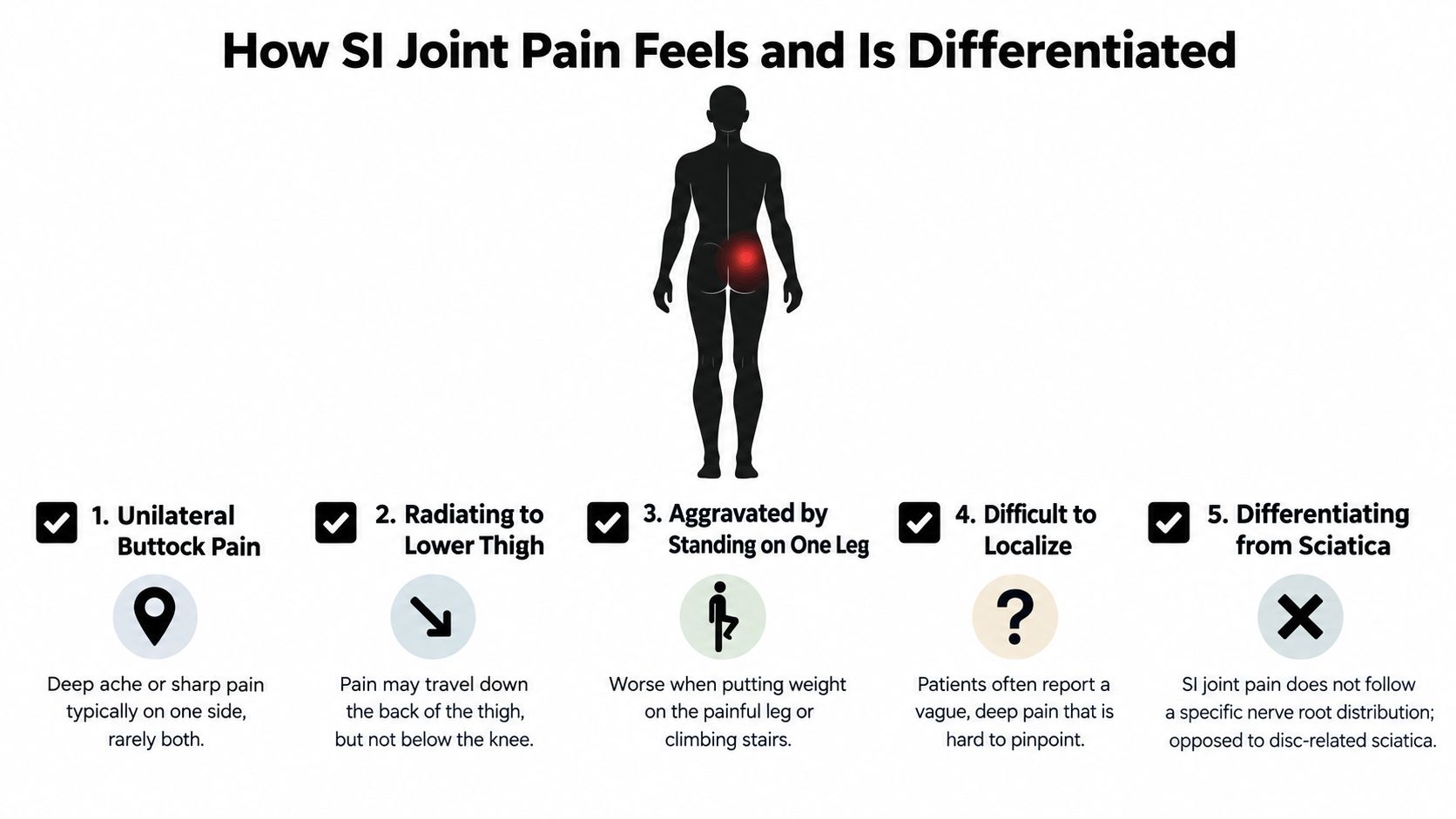
SI joint pain often appears below the beltline, slightly to one side of the lower back, near the upper buttock. It may spread toward the hip or thigh and can resemble lumbar disc pain, hip pain, or sciatica. The location helps, but the behavior of the pain often provides more useful clues.
Common aggravating activities include prolonged sitting, standing, stair climbing, single-leg loading, and transitional movements such as rising from a chair. Because several conditions can produce the same pattern, self-diagnosis isn't reliable.
What the examination is trying to reproduce
A clinician doesn't rely on one maneuver. The examination may include:
- Thigh thrust: Loads the pelvis through the femur to see whether it reproduces the familiar SI-region pain.
- FABER or Patrick test: Places the hip in flexion, abduction, and external rotation, helping assess the SI region and nearby hip structures.
- Gaenslen test: Positions the legs to stress the pelvis and SI joints.
- Distraction and compression: Applies controlled force to the pelvic ring from different directions.
- SI shear testing: Challenges the joint and surrounding stabilizers with a shearing force.
Evidence-based literature recommends at least three positive provocative maneuvers before radiologically controlled infiltration testing. The IASP criteria require pain in the SI region, reproduction of that pain with SI-selective clinical tests, and complete relief after selective joint infiltration (evidence-based SI joint diagnosis).
What happens after the examination
The clinician also considers lumbar nerve symptoms, hip range of motion, strength, sensation, reflexes, gait, prior surgery, and inflammatory features. If the findings point toward the SI joint, a fluoroscopy-guided injection may help confirm the source and, when appropriate, provide therapeutic relief.
A diagnostic procedure should answer a specific question. It shouldn't be used solely because a scan is normal or because pain happens to be located near the pelvis.
Next Steps for Lasting Relief in the Chicago Ridge Area
Cause identification changes the plan. A patient with a recent strain may need activity modification and a carefully supervised rehabilitation strategy. Someone with inflammatory features may need evaluation for an underlying inflammatory condition. A person with persistent pain after fusion may need a broader review of altered biomechanics, adjacent-segment problems, and other post-surgical pain sources.
The first step is a focused history. Bring details about when the pain began, whether there was a fall or collision, how sitting and stair climbing affect it, whether pregnancy or delivery preceded the symptoms, and whether you have had lumbar surgery. Also mention psoriasis, inflammatory arthritis, infection risk, cancer history, leg length concerns, or a sustained change in walking pattern.
Choosing an appropriate evaluation
A pain and wellness clinic can coordinate several parts of the assessment while keeping the suspected pain generator in view. Depending on the findings, care may include:
- Conservative support: Activity changes, medication discussion, stabilization strategies, and coordination with rehabilitation professionals.
- Diagnostic intervention: A selective, image-guided SI joint injection when examination findings support that question.
- Multimodal treatment: Interventional procedures combined with appropriate rehabilitation, primary care coordination, or surgical consultation.
- Post-surgical review: A careful assessment when pain continues after lumbar fusion, rather than assuming the original spinal diagnosis explains every symptom.
Reviews report SI-joint prevalence of 32% to 37% after lumbosacral fusion, and the SI joint was identified as the pain source in about 40% of patients with residual off-center low back pain after lumbar fusion (review of SI joint pain after lumbar fusion). Those findings support evaluating the SI joint when pain persists after surgery, while still considering other possible causes.
Midwest Pain & Wellness is a Chicago Ridge pain and wellness clinic, not a physical therapy practice. Led by double board-certified interventional pain specialist Dr. Yaw Donkoh, the clinic provides opioid-sparing, multimodal care and coordinates with primary care providers, surgeons, chiropractors, and rehabilitation therapists for patients in Chicago Ridge, Palos Heights, Palos Hills, Evergreen Park, and Hickory Hills, Illinois. You can review sacroiliac joint pain treatment options to understand how evaluation and image-guided care may fit into a broader plan.
Don't wait indefinitely if pain is limiting walking, sleep, work, or basic transitions. Seek prompt medical evaluation for severe or rapidly worsening pain, fever, new weakness, bowel or bladder changes, unexplained weight loss, or pain after significant trauma.
If pain near your buttock or lower back has persisted, Midwest Pain & Wellness can evaluate whether the SI joint, surrounding tissues, lumbar spine, or another structure is contributing. Visit Midwest Pain & Wellness to request an appointment in Chicago Ridge, Illinois, and discuss a personalized, opioid-sparing plan for restoring function.