You've been dealing with a nagging ache just below your belt line. It's worse when you climb stairs, roll onto one side in bed, or stand up after sitting, yet your back imaging hasn't given you a clear explanation. Sometimes the pain travels into the buttock or back of the thigh, which can make it feel like sciatica. That pattern is common with sacroiliac joint pain, but it isn't specific enough for self-diagnosis.
The sacroiliac joint can produce real, persistent pain while remaining difficult to identify from symptoms alone. Understanding where the pain is located, what movements reproduce it, and how it differs from nerve, disc, hip, or facet pain can make your clinical evaluation more productive.
Table of Contents
- What Is Sacroiliac Joint Pain and Why It Matters
- Common Sacroiliac Joint Pain Symptoms and Patterns
- Distinguishing SI Joint Pain from Other Conditions
- How SI Joint Pain Is Diagnosed
- Treatment Options for Sacroiliac Joint Pain
- Common Misconceptions About SI Joint Pain
- When to Seek Urgent Evaluation for Back and Pelvic Pain
- Next Steps for Managing Sacroiliac Joint Pain
What Is Sacroiliac Joint Pain and Why It Matters
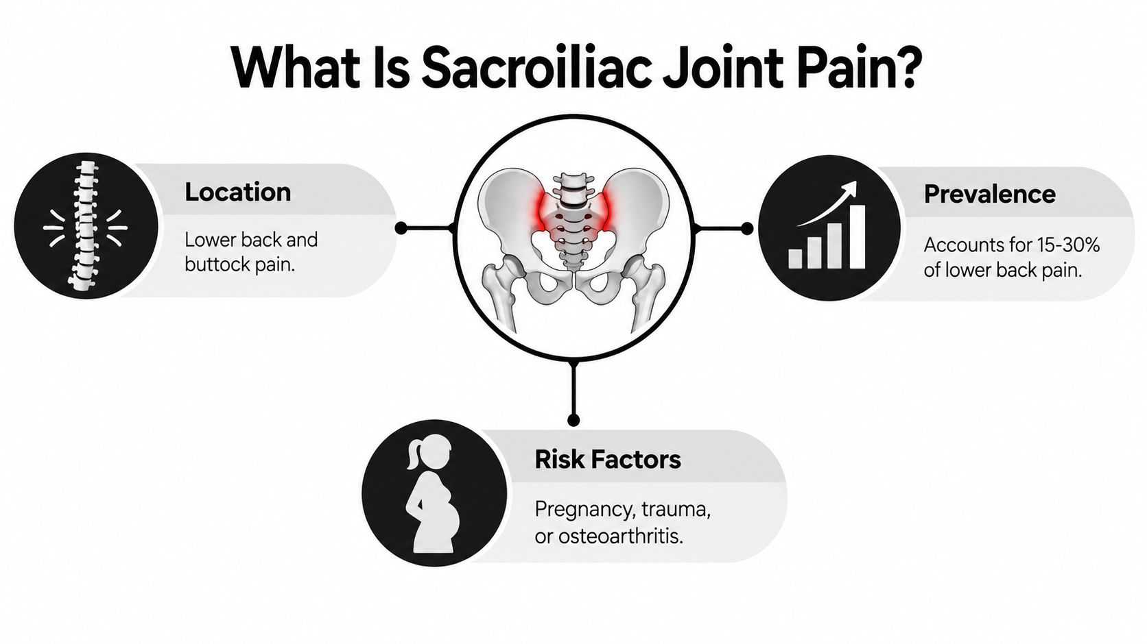
A patient may point to one spot beside the lower spine and say, “That's where it starts.” The discomfort often feels deep rather than superficial, and it may spread into the buttock or the back of the thigh. Sitting through a drive, shifting position in bed, or putting more weight on one leg can bring it on.
The sacroiliac joints sit where the sacrum, the base of the spine, meets the ilium, the broad part of the pelvis. They transfer load between your upper body and legs during walking, standing, sitting, and transitions. Although the joints move only slightly, they're exposed to repeated forces and can become painful after trauma, repetitive strain, pregnancy-related pelvic changes, or altered mechanics after surgery or injury.
Why the source is easy to miss
Sacroiliac joint pain is not a rare diagnosis. A peer-reviewed review estimates that it accounts for approximately 15% to 30% of chronic, non-radicular low-back-pain cases (peer-reviewed review). “Non-radicular” means the pain doesn't primarily result from a compressed spinal nerve root.
That doesn't mean every one-sided buttock ache comes from the SI joint. A disc problem, facet joint pain, hip disorder, pelvic condition, or nerve irritation can create similar symptoms. No single symptom proves the SI joint is responsible, so a careful assessment must compare competing explanations.
Clinical perspective: The location of pain gives us a starting point. The pattern of provocation, examination findings, and response to a properly performed diagnostic procedure provides stronger evidence.
People often search for general strategies for joint discomfort while deciding whether to seek care. A resource on gentle arthritis pain relief may offer broad self-care context, but persistent or function-limiting SI-region pain still deserves an evaluation matched to your spine, hip, pelvis, and nerves.
Common Sacroiliac Joint Pain Symptoms and Patterns
The most recognizable pattern is deep, one-sided pain just below and slightly toward the middle from the posterior superior iliac spine, or PSIS. The PSIS is the bony prominence you can feel on either side of the lower back. Many people call this area the “back pocket” region.
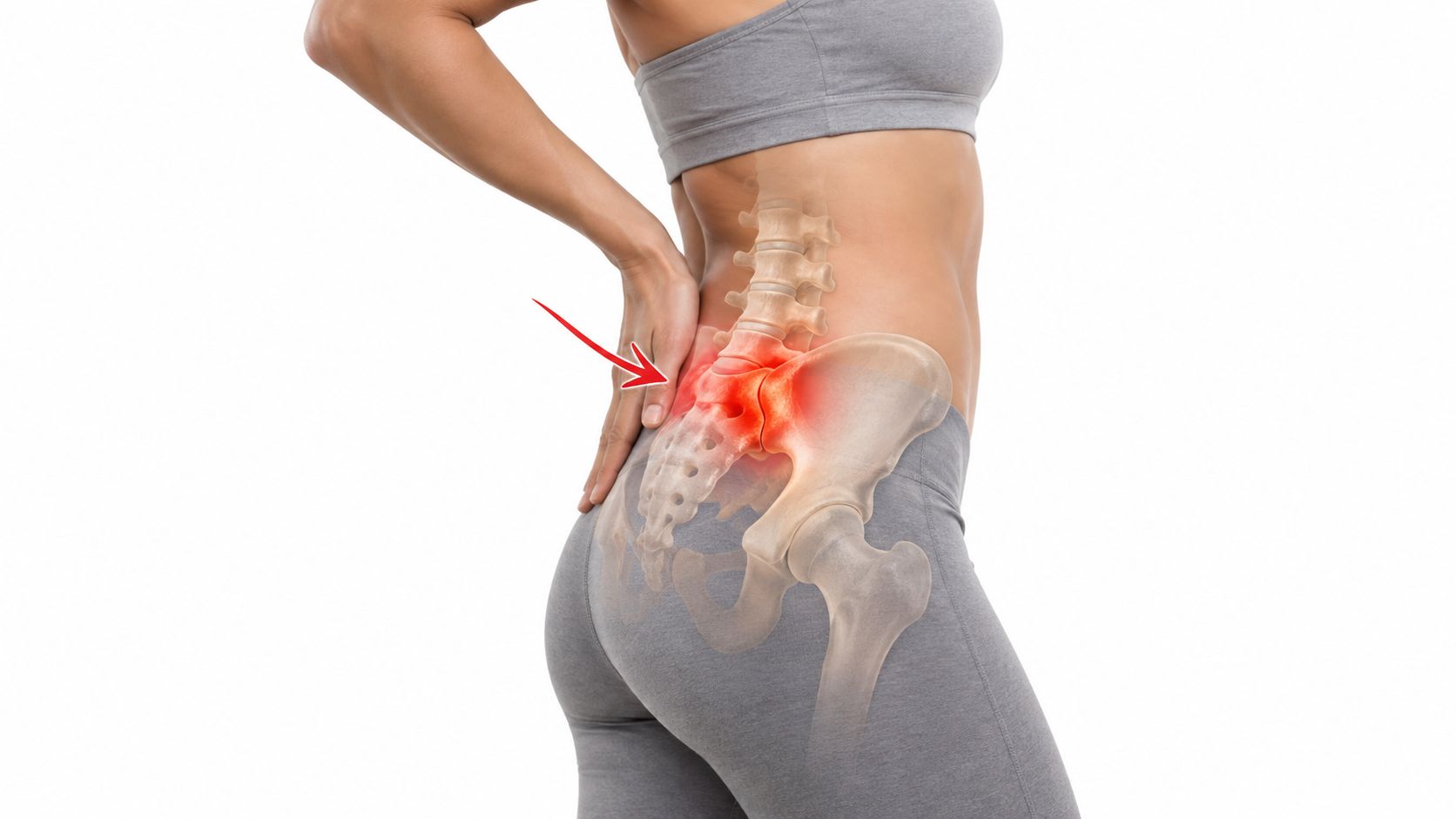
Pain may remain near the joint or refer through the buttock and down the back of the thigh, usually reaching around the knee. It can also be felt toward the hip, groin, or another part of the lower extremity. The clinical description of SI joint symptoms emphasizes that these patterns are useful clues, not diagnostic proof.
Mechanical triggers matter
SI joint symptoms often change with load and position. Common aggravating activities include:
- Prolonged sitting: Remaining in one position can increase pressure around the joint.
- Rolling or lying on the painful side: Direct compression may reproduce familiar pain.
- Stair climbing: Alternating single-leg loading can stress the painful region.
- Transfers: Moving from sitting to standing or getting out of a vehicle may trigger a sharp catch.
- Asymmetric walking: A limp or repeatedly favoring one leg can intensify symptoms.
Some patients describe clicking, catching, pelvic instability, or a feeling that the pelvis is “out.” Others notice gait awkwardness without obvious clicking. These details help a clinician understand how the pain behaves, but hip, lumbar, pelvic, and neurologic disorders can create the same sensations.
A useful distinction is whether the examination reproduces your familiar pain, rather than causing pressure or discomfort somewhere in the pelvis. A maneuver that hurts but feels unrelated to your usual symptoms provides weaker evidence than one that recreates the exact complaint.
Distinguishing SI Joint Pain from Other Conditions
SI joint pain is often mistaken for sciatica because both conditions can travel into the buttock or leg. Prevalence estimates vary according to the population and diagnostic method, with SI joint pain estimated to account for 10% to 38% of low-back-pain cases (clinical review). Reported clinical prevalence has varied widely, which reflects how differently clinicians define and confirm the diagnosis.
The comparison below shows the tendencies clinicians consider. These are patterns, not rules that can replace an examination.
| Condition | Pain location | Radiation pattern | Aggravating factors | Distinguishing features |
|---|---|---|---|---|
| SI joint pain | Usually below the belt line and beside the lower spine, often in the buttock | Buttock and posterior thigh, sometimes toward the groin or lower extremity; below-knee radiation is less common | Sitting, lying on the painful side, stairs, transfers, asymmetric weight-bearing | Familiar pain reproduced by a cluster of SI provocation tests |
| Lumbar disc pain | Central or slightly off-center low-back pain | May extend into the leg, sometimes following a nerve distribution | Sitting, bending, lifting, or spinal loading may aggravate it | Disc findings must correlate with symptoms and examination |
| Lumbar facet pain | More central or paraspinal low-back pain | May refer into the buttock or thigh | Extension, rotation, and prolonged standing may be provocative | Pain is associated with facet loading, but no single maneuver confirms the source |
| Hip osteoarthritis or other hip pathology | Often centered in the groin, front of the hip, or deep joint | May refer to the thigh or knee | Walking, pivoting, and hip movement | Restricted hip motion, especially internal rotation, may be important |
| Lumbar radiculopathy | Back pain with a more distinct nerve-related leg pattern | Sharp or electric pain may travel below the knee | Coughing, straining, certain spinal positions, or nerve tension | Numbness, weakness, reflex changes, or other neurologic findings can occur |
A patient can also have more than one pain generator. That's why an evaluation at a clinic offering lumbar pain management should examine the lumbar spine, hips, neurologic system, and SI region rather than treating every buttock symptom as one condition.
How SI Joint Pain Is Diagnosed
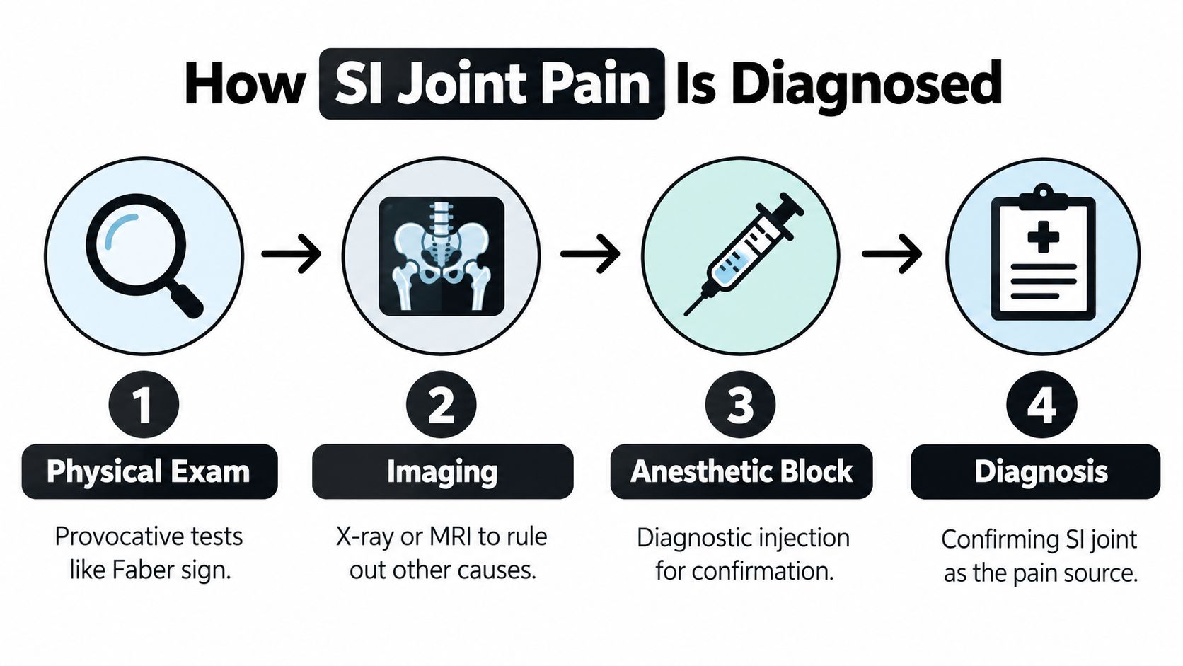
Diagnosis begins with the story. A clinician will ask where the pain started, whether there was a fall or lifting injury, how pregnancy or surgery may have changed your mechanics, and which movements reproduce the complaint. Gait, posture, hip motion, lumbar movement, strength, sensation, and reflexes may all contribute to the assessment.
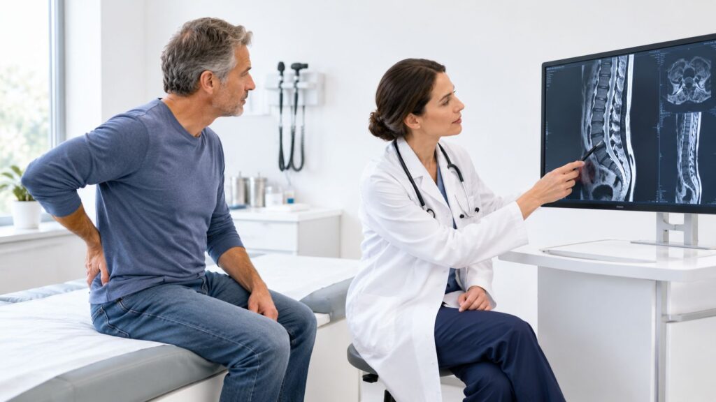
Imaging has a supporting role. X-rays or MRI can help identify fractures, significant hip or lumbar disease, inflammatory changes, or other concerning conditions, but an imaging finding doesn't automatically identify the painful structure. Age-related or incidental changes can appear without being the cause of your symptoms.
Why clinicians use a test cluster
The examination commonly includes:
- Gaenslen test: Places stress across the pelvis through different hip positions.
- Thigh-thrust test: Applies a controlled force through the femur toward the SI joint.
- Distraction test: Applies outward pressure across the front of the pelvis.
- Compression test: Loads the pelvis from the sides.
- Sacral-thrust test: Applies pressure through the sacrum.
FABER, which combines hip flexion, abduction, and external rotation, may also help assess the SI region and hip. A diagnostic accuracy review found variable performance among individual maneuvers. The thigh-thrust test was the most sensitive at 74.4%, while FABER was the most specific at 66.7% (diagnostic accuracy review).
The practical rule is that at least three positive provocation tests, supported by the patient's familiar pain and the rest of the examination, increase confidence that the SI joint may be involved. A single positive test is much less persuasive because several maneuvers can stress neighboring structures.
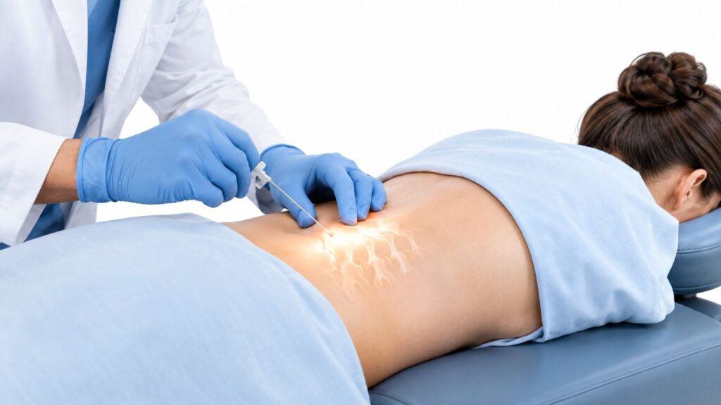
When uncertainty remains, an image-guided anesthetic injection can provide additional confirmation. A clinician places medication into the SI joint while using imaging to guide needle position. Meaningful, temporary relief during the expected anesthetic period supports the joint as a pain source, though the result still needs to fit the broader clinical picture. Patients can learn more about the role of a fluoroscopy-guided injection before discussing whether it's appropriate.
Treatment Options for Sacroiliac Joint Pain
Treatment should follow the diagnosis, not the other way around. If the examination points toward the SI joint but also suggests hip or lumbar involvement, the plan may need to address more than one contributor.
Early care often includes activity modification, a targeted rehabilitation program, and medication when medically appropriate. Physical therapy may focus on pelvic control, trunk and hip strength, movement coordination, and gradual return to activities. A brace can help selected patients, but it isn't a universal solution, and exercises that aggravate familiar pain should be adjusted rather than forced.

When targeted procedures make sense
If conservative care hasn't provided enough improvement, an image-guided SI joint injection may serve two purposes. The anesthetic can help test whether the joint contributes to the pain, while a corticosteroid may reduce inflammation and provide therapeutic relief. The benefit can be temporary, and the procedure shouldn't be used as a substitute for correcting movement, strength, or workload factors that continue to irritate the region.
Patients comparing at-home approaches may also review a Granted Solutions buyer guide for general information about pain-relief devices. Such products may complement a plan, but they can't determine whether pain comes from the SI joint, lumbar spine, hip, or a nerve.
For patients who receive meaningful but temporary relief from diagnostic blocks, radiofrequency ablation may be discussed. This procedure interrupts pain signals carried by selected lateral sacral branches. It doesn't repair every structural problem, and candidacy depends on the diagnostic response, examination, medical history, and procedural goals.
Other options, including peripheral nerve stimulation, spinal cord stimulation, or regenerative treatments such as PRP, may be considered selectively. Cell-based therapies remain investigational for most spine applications. The treatment pathway at Midwest Pain & Wellness for SI joint pain can be discussed in the context of a personalized, opioid-sparing plan.
Common Misconceptions About SI Joint Pain
A common reason SI joint pain gets missed is that it can look like several other problems at once.
Myth: Pain near the belt line must come from a disc.
Pain in this area can come from the lumbar discs, but the SI joint often causes a deep ache low in the back or buttock, usually more on one side. Some patients also feel pain into the groin, outer hip, or upper thigh. If the pattern is assumed to be a disc problem from the start, the exam and treatment plan can drift away from the actual pain source.
Myth: Normal imaging rules out SI joint pain.
It does not. X-rays, CT, and MRI are useful because they help rule out fractures, inflammatory disease, hip problems, or spine conditions that need a different workup. But many patients with SI-mediated pain do not have one clear imaging finding that settles the diagnosis, which is why symptom pattern and physical examination matter.
Myth: A positive provocation test confirms the diagnosis.
Provocation tests are helpful when they reproduce the patient's familiar pain, especially as part of a cluster, but they are not definitive on their own. A 2021 systematic review and meta-analysis found pooled sensitivity of 0.83 and specificity of 0.59, with a false-positive rate of 0.41. The same review estimated only 35% certainty that a positive cluster correctly identified the SI joint as the pain generator. In practice, that means these tests guide diagnostic reasoning. They do not replace it.
Myth: SI joint pain automatically means surgery.
Treatment usually starts with conservative care, then moves to targeted procedures only when the history, exam, and response to treatment support that choice.
Pregnancy, trauma, and repeated mechanical strain can all precede symptoms. Persistent pain after those events deserves a careful evaluation, not dismissal.
When to Seek Urgent Evaluation for Back and Pelvic Pain
A flare of pain near the SI joint is often mechanical and can wait for an outpatient visit. Some symptom patterns should not wait, because they can point to a nerve emergency, infection, fracture, inflammatory disease, or another condition that needs faster workup.
Get urgent medical care if you have any of the following:
- Progressive leg weakness or loss of coordination: This raises concern for meaningful nerve involvement.
- New bowel or bladder dysfunction: Trouble controlling or emptying the bladder or bowel needs immediate assessment.
- Saddle numbness: Numbness around the groin, inner thighs, or seat area is an emergency warning sign.
- Fever, unexplained weight loss, or systemic illness: Back or pelvic pain with these features can signal infection, malignancy, or another serious illness.
- Severe, unrelenting pain: Pain that keeps escalating or does not ease with usual measures deserves prompt evaluation.
A different history can point away from routine mechanical SI pain. If pain began before age 45, developed gradually, has lasted for months, improves with movement more than rest, or wakes you at night and eases after getting up, I start thinking about inflammatory sacroiliitis or axial spondyloarthritis rather than a simple strain. In that setting, imaging may be used to look for active inflammation, but the symptom pattern still matters.
Without these red flags, do not ignore persistent symptoms. Schedule an evaluation if pain keeps returning, limits walking, interrupts sleep, makes sitting or standing difficult, or stops you from working and exercising normally. An interventional pain specialist can examine the lumbar spine, hip, pelvis, and nerves together, then decide whether diagnostic injection, rehabilitation, imaging, or referral is the next reasonable step.
Next Steps for Managing Sacroiliac Joint Pain
Start by recording the pattern rather than only the intensity. Note whether pain is below the belt line, whether it stays on one side, how far it travels, and which activities reproduce your familiar symptoms. Include any fall, lifting event, pregnancy-related change, surgery, or altered gait that preceded the problem.
Bring that information to a clinician who evaluates the lumbar spine, hip, pelvis, and nervous system together. SI joint pain is commonly considered through a cluster of provocation findings, not one symptom or scan. If the diagnosis remains uncertain, an image-guided anesthetic block may help determine whether the joint is a meaningful pain generator.
For residents of Chicago Ridge, Palos Heights, Palos Hills, Evergreen Park, and Hickory Hills, Illinois, Midwest Pain & Wellness provides interventional pain evaluation under double board-certified interventional pain specialist Dr. Yaw Donkoh. Care may include targeted physical therapy, SI joint injections, radiofrequency ablation, and other image-guided or multimodal treatments selected according to the diagnosis and response to earlier care.
The practical objective isn't merely to quiet pain for a short period. It's to improve walking, sitting, sleep, work, and daily movement while reducing reliance on opioids. Persistent sacroiliac joint pain symptoms deserve a clear explanation and a plan that matches the actual pain source.
Midwest Pain & Wellness evaluates sacroiliac joint pain symptoms and offers image-guided SI joint injections, radiofrequency ablation, targeted rehabilitation, and other opioid-sparing options when appropriate. If pain below your belt line or in your buttock is limiting normal activity, visit Midwest Pain & Wellness to request an evaluation in Chicago Ridge, Illinois.