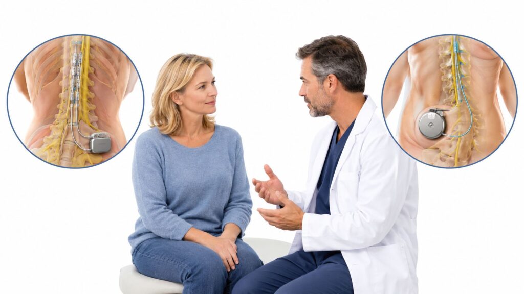
You've had aching low-back pain for weeks or months. It sits just below the beltline, close to one buttock, and flares when you stand too long, climb stairs, walk, or roll over in bed. Then your lumbar MRI comes back without a clear explanation, leaving you wondering whether the pain is real, whether something was missed, or whether the problem might be coming from the sacroiliac joint.
That uncertainty is common. SI joint pain diagnosis isn't based on one symptom, one scan, or one physical maneuver. At a pain and wellness clinic, the evaluation usually develops in stages: a careful history, examination with provocation tests, selective imaging when needed, and, when uncertainty remains, an image-guided diagnostic injection. Midwest Pain & Wellness in Chicago Ridge, Illinois, serves patients from Palos Heights, Palos Hills, Evergreen Park, and Hickory Hills with structured, opioid-sparing pain care.
Table of Contents
- Why Your Low-Back Pain Might Be Your SI Joint
- How the SI Joint Works and Why It Hurts
- What Your Medical History Reveals About SI Joint Pain
- Physical Provocation Tests and What They Really Tell Us
- Why Imaging Alone Cannot Confirm SI Joint Pain
- The Diagnostic SI Joint Injection Explained
- Ruling Out Other Pain Sources and Coordinating Your Care
- Your Next Steps for SI Joint Pain Evaluation
Why Your Low-Back Pain Might Be Your SI Joint
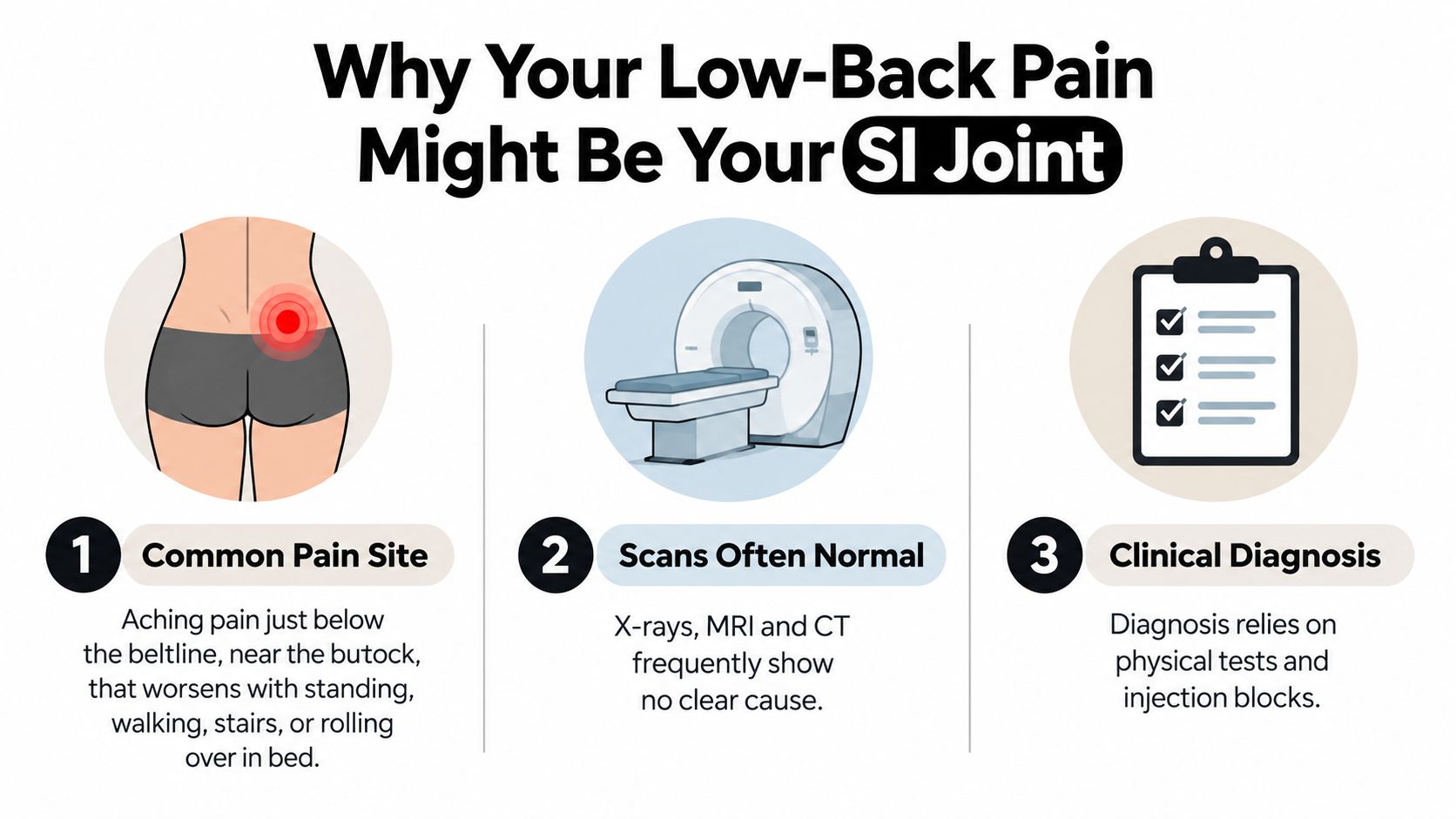
A patient may point to pain beside the low back, near the dimple above one buttock. Standing at the kitchen counter brings it on. A grocery-store walk, stairs, or turning in bed makes it worse, yet a lumbar scan shows no clear disc problem. That pattern makes the SI joint worth investigating, but it does not identify the source by itself.
The sacroiliac joint can mimic several conditions, including lumbar disc or facet pain, hip disease, muscle injury, and nerve irritation. Pain often sits below the fifth lumbar vertebra and near the buttock. Symptoms may increase with standing, walking, climbing stairs, or changing position. These clues help guide the examination, like labels on a map, but they cannot confirm the destination. A careful evaluation compares the SI joint with other plausible pain sources.
Why clinicians take the possibility seriously
The SI joint is a reasonable suspect, not an automatic answer. A systematic review estimates that SI joint pain accounts for roughly 10% to 25% of persistent axial low-back pain after certain disc and nerve-related conditions are excluded (systematic review of SI joint pain prevalence). Estimates vary widely, from 10% to 62%, because studies use different patient groups, referral settings, exclusion rules, and reference tests. The broad range is a reason to interpret prevalence cautiously, not to assign a diagnosis from a percentage.
That distinction matters in Illinois pain care. A reassuring lumbar scan does not close the case, and a positive provocation maneuver does not settle it. The usual pathway builds evidence in stages: history, a cluster of examination findings, selective imaging, and, when appropriate, an interpreted image-guided diagnostic injection. Midwest Pain & Wellness in Chicago Ridge, Illinois, serves patients from Palos Heights, Palos Hills, Evergreen Park, and Hickory Hills with structured, opioid-sparing pain care.
People who spend long periods seated can also review Pittsburgh back care tips from Full Circle. Those ideas may support general back care, but persistent pain, spreading symptoms, or difficulty walking and sleeping calls for clinical evaluation.
How the SI Joint Works and Why It Hurts
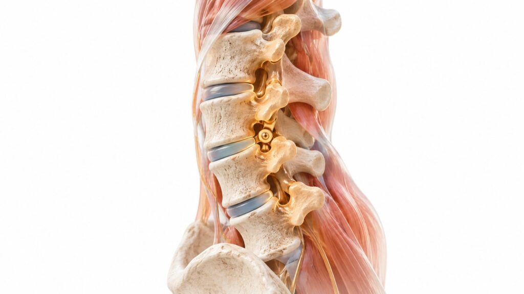
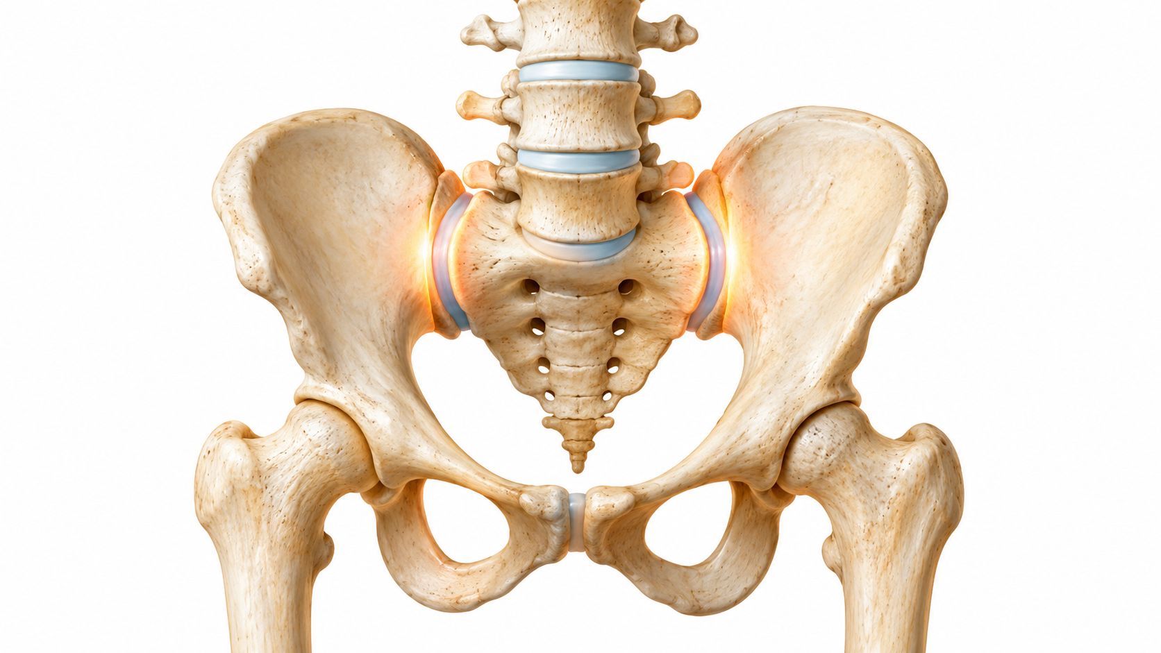
You have an SI joint on each side, where the triangular sacrum at the base of the spine meets the pelvic bones. Each joint functions as a load-transfer point and stabilizer. It passes force between your upper body and legs as you stand, walk, climb stairs, or change position.
The joint moves only slightly, yet that small motion has an important job. If force is distributed unevenly through the pelvis, repeated stress can irritate the joint surfaces, supporting ligaments, or nearby tissues. A structure that barely moves can still produce persistent pain when it absorbs load with every step.
How the joint becomes overloaded
Several situations can raise stress around the SI region:
- Degenerative change: Wear in the joint or nearby structures may change how forces are distributed.
- Ligament strain: A fall, twisting injury, or repeated uneven movement can irritate stabilizing tissues.
- Pregnancy-related loosening: Changes in the pelvic ligaments may contribute to pain during or after pregnancy.
- Prior lumbar fusion: Altered spinal mechanics can send more force toward the SI joints.
- Trauma: A direct impact or uneven loading event may trigger one-sided symptoms.
Pain can stay near the buttock or spread into nearby areas. This is referred pain. It does not necessarily indicate nerve compression, and its location alone cannot confirm the SI joint as the source.
The same buttock ache can come from the lumbar spine, hip, pelvic muscles, or other tissues. Because prevalence estimates shift with referral patterns and study design, the anatomy and load mechanics described here matter more than any single percentage (evidence on SI joint prevalence and diagnostic difficulty). Positive examination maneuvers can screen for an SI-region pattern, but they do not settle the diagnosis by themselves.
A useful mental model
The pain generator may be inside the joint itself or in the surrounding ligaments and posterior structures. These sources can feel similar while requiring different evaluation and treatment. Load-related irritation is therefore the more useful lens: the next step is a structured history that connects symptoms with movement, injury, pregnancy, surgery, and other possible sources.
What Your Medical History Reveals About SI Joint Pain
The first appointment usually begins before anyone performs a maneuver. I want to know exactly where the pain starts, where it travels, what brings it on, and what makes it settle. The details help separate an SI-region pattern from symptoms more suggestive of a lumbar disc, hip, or nerve problem.
A useful description includes the side of the pain, its location relative to the beltline, whether it reaches the groin or leg, and whether it feels aching, sharp, burning, or electric. Tell your clinician whether standing, walking, stairs, getting out of a chair, or rolling in bed changes the pain. Also mention prior falls, pregnancy, lumbar surgery, hip disease, inflammatory conditions, and treatments that have or haven't helped.
Useful symptom cluster: Pain below the fifth lumbar vertebra, often near the buttock, that increases with standing, walking, stair climbing, or position changes raises suspicion for SI-region pain, but it doesn't confirm the source.
Red flags come first
A pain specialist also screens for signs that could point to infection, cancer, fracture, serious inflammation, or neurologic compromise. Report fever, unexplained weight loss, new or progressive weakness, significant numbness, saddle-area sensory changes, or bowel and bladder changes promptly. These findings don't automatically identify a particular disease, but they can change the urgency and direction of the evaluation.
Bring prior imaging reports and, if available, the actual images. A medication list, surgical history, injection history, and short pain diary can make the visit more productive. Note when symptoms occur rather than relying on a general statement such as “my back always hurts.”
Your history also helps determine whether imaging is necessary and which region deserves attention. If pain is strongly linked to hip motion, the hip may need evaluation. If burning pain travels with numbness or weakness, the lumbar nerves deserve closer review. The interview doesn't replace the examination. It tells the clinician how to interpret it.
Physical Provocation Tests and What They Really Tell Us
Provocation tests are designed to load the SI region and see whether they reproduce the patient's familiar pain. The commonly used group includes Gaenslen, thigh thrust, distraction, compression, and sacral thrust. Some clinicians also use FABER or SI shear maneuvers as part of a broader examination.
Here is what the standard tests generally do:
| Test | What Happens | What It Stresses |
|---|---|---|
| Gaenslen | One hip is flexed while the other leg extends, creating an asymmetrical pelvic position. | SI-region structures under torsional load |
| Thigh thrust | The clinician applies a controlled force through the thigh while the patient lies on the back. | One side of the pelvis and SI joint |
| Distraction | Pressure is applied to the front of the pelvic bones, gently spreading them. | Anterior SI-region structures |
| Compression | Pressure is applied to the sides of the pelvis. | Posterior pelvic and SI-region structures |
| Sacral thrust | A controlled force is applied through the sacrum. | SI-region structures through the sacral base |
A positive cluster is not a verdict
Many patient-facing pages imply that three positive tests establish SI joint pain. They don't. A cluster of at least three positive maneuvers can increase suspicion and help direct the next step, but physical testing alone has limited rule-in accuracy.
A 2021 systematic review and meta-analysis reported a positive likelihood ratio of 2.13, a negative likelihood ratio of 0.33, and a diagnostic odds ratio of 9.01 for clusters of provocation tests (systematic review of SI joint provocation-test clusters). Assuming a 20% underlying prevalence, a positive cluster raised the estimated probability to only 35%, while a negative cluster reduced it to approximately 8%.
That pattern matters. These tests are generally more useful for lowering suspicion when the cluster is negative than for proving the SI joint is the pain generator when the cluster is positive.
Practical rule: Three or more positive tests support the question, “Should we investigate the SI joint further?” They don't answer, “Has the SI joint definitely caused this pain?”
The examination must still consider lumbar disc and facet disorders, hip pathology, neurologic findings, pelvic problems, and myofascial pain. A stiff hip or degenerative lumbar spine can make several pelvic maneuvers uncomfortable without making the SI joint the primary source. Evidence supporting provocation clusters is also considered very low certainty, so the findings belong within the complete clinical picture (primary-care guidance on SI joint diagnosis).
Why Imaging Alone Cannot Confirm SI Joint Pain
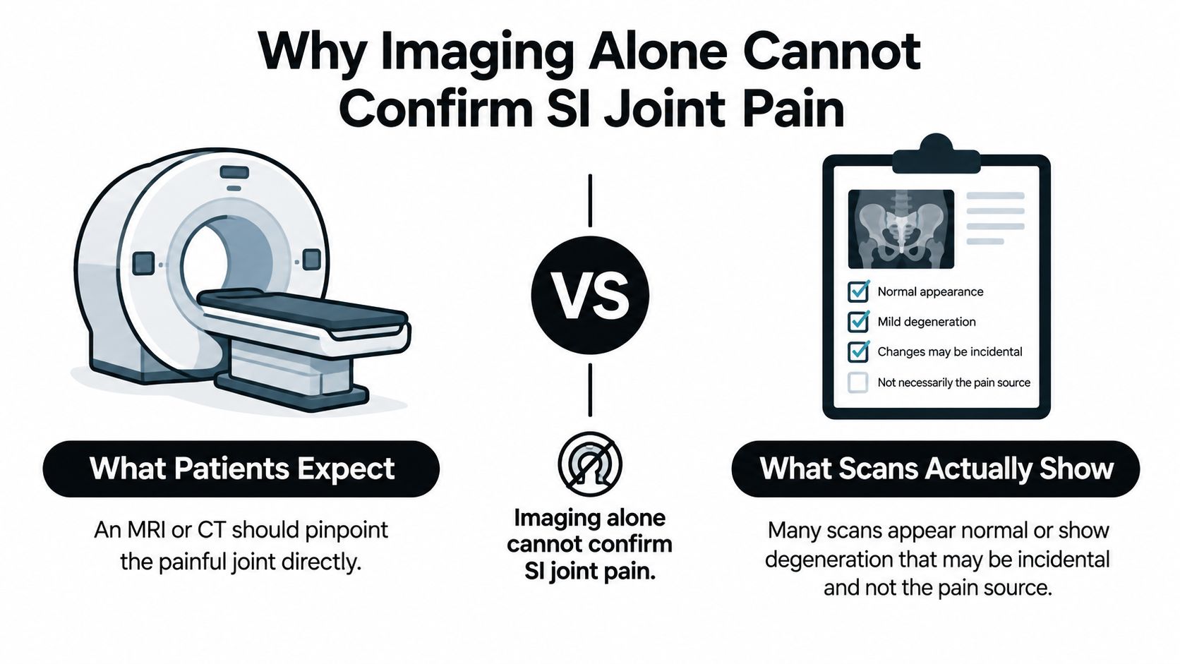
Patients often expect an MRI or CT scan to point directly to the painful SI joint. That expectation is understandable, but imaging answers a different question. It can reveal structural changes and help identify dangerous or competing conditions, yet it usually can't prove that an SI joint abnormality is causing the patient's symptoms.
A scan may help detect or exclude fracture, inflammatory sacroiliitis, infection, malignancy, or hip disease. It may also show degenerative changes. Those findings can be important, but degeneration alone doesn't establish that the SI joint is the active pain generator.
What a normal scan means
A normal-looking SI joint doesn't mean the pain is imaginary. SI joint pain commonly lacks consistent radiographic or laboratory abnormalities. Conversely, an abnormal-looking joint may not be responsible for the symptoms.
| Imaging finding | What it can contribute | What it cannot establish |
|---|---|---|
| Normal-appearing SI region | Makes some structural conditions less apparent. | It cannot rule out clinically meaningful SI-region pain. |
| Degenerative SI changes | Shows wear or structural alteration. | It cannot prove those changes cause the pain. |
| Inflammatory features | May support further evaluation for inflammatory disease. | It doesn't replace clinical correlation. |
| Hip or lumbar abnormality | May identify another possible pain source. | It doesn't automatically exclude SI-region involvement. |
A scan is most useful when its findings match the history and examination. If the pain pattern points toward the SI region but imaging shows only mild lumbar wear, the clinician may continue the structured evaluation rather than treating the scan as the final answer. Patients who want a broader explanation of how clinicians approach back-pain workups can review this guide to diagnosing back pain.
The key distinction is simple: imaging shows anatomy; diagnosis identifies the structure producing the familiar pain. Those are related, but they aren't identical.
The Diagnostic SI Joint Injection Explained
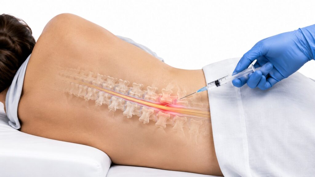
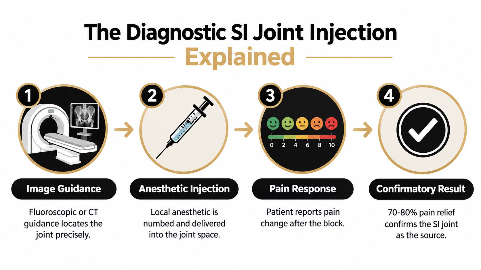
You may leave an appointment with several clues pointing toward the SI joint, yet still lack a definitive answer. When the history and examination make it a leading possibility, an image-guided intra-articular local-anesthetic block can test whether numbing that joint changes your familiar pain. It is a controlled experiment, not a perfect oracle.
The procedure usually follows these steps:
- Positioning and preparation: You are positioned for access to the SI joint. The skin is cleaned, and the area is numbed.
- Image guidance: Fluoroscopy or CT guides the needle into the joint.
- Pain reproduction: The clinician notes whether needle placement or injection reproduces your usual pain.
- Anesthetic response: You report how the familiar pain changes while the local anesthetic is active.
- Documentation: Pain levels and relevant activities are recorded so the result can be interpreted rather than guessed.
What the injection is testing
The clinician is asking two questions. Did entering and injecting the joint reproduce the pain that brought you in? Did meaningful relief follow during the period when the anesthetic should be working?
A useful result means improvement in the familiar, function-limiting pain, not merely temporary comfort near the needle. You may be asked to perform safe activities that normally trigger symptoms and record what happens afterward. That information gives the response context.
Relief thresholds vary by protocol. Dual controlled blocks commonly require at least 70% pain relief, single blocks often use at least 75%, and a response between roughly 50% and 75% may indicate that the SI joint contributes substantially without proving it is the only source (review of diagnostic SI joint blocks). These thresholds are not interchangeable, so your clinician should explain which protocol applies.
A single uncontrolled block has an estimated false-positive rate of about 20%, which helps explain why comparative or dual blocks provide stronger diagnostic support (diagnostic accuracy review). Pain relief can result from medication spreading outside the joint, expectation, or changes in activity during the observation period. A positive injection therefore strengthens the diagnosis, but it does not erase the need to consider other pain sources.
In the Chicago Ridge area, the visit generally includes preparation, image-guided needle placement, brief observation, and instructions for tracking your response. Details depend on the procedure and your medical history. You can review information about fluoroscopy-guided injections before discussing whether this test fits your evaluation.
Ruling Out Other Pain Sources and Coordinating Your Care
SI joint diagnosis is strongest when several pieces point in the same direction. A clinician compares the symptom pattern, examination findings, imaging, and injection response rather than allowing one positive maneuver or one scan to dominate the decision.
Common look-alikes
- Lumbar disc or facet pain: The lumbar examination and neurologic assessment may reveal patterns that fit the spine more closely.
- Hip arthritis: Groin pain, restricted hip motion, and pain reproduced through the hip may redirect the workup.
- Radiculopathy: Burning or electric pain with numbness, weakness, or reflex changes can suggest nerve-root involvement.
- Piriformis or myofascial pain: Local muscle tenderness and pain with specific movements may point to soft tissues around the pelvis.
- Inflammatory sacroiliitis: Morning stiffness, systemic symptoms, or inflammatory features may require medical evaluation beyond a mechanical pain workup.
Blood tests may be considered when the history raises concern for inflammation or systemic disease, but a normal result doesn't by itself diagnose or exclude mechanical SI-region pain. Patients who want plain-language background on inflammation testing can review this inflammation panel guide.
Inside the joint or around it
The SI region includes more than the articular surfaces. Pain can arise from extra-articular ligaments and posterior-complex tissues, and those sources may imitate pain from inside the joint. Distinguishing them matters because a treatment aimed at intra-articular pain may not address ligamentous pain.
Care coordination helps prevent repeated, disconnected testing. A pain and wellness clinic may review records from a primary care provider or surgeon, communicate with chiropractors and rehabilitation therapists, and use the findings to build a multimodal plan. Around Chicago Ridge, Palos Heights, Palos Hills, Evergreen Park, and Hickory Hills, patients may already have several clinicians involved. The goal is to give each clinician the same diagnostic picture, not to repeat the same evaluation in isolation.
Midwest Pain & Wellness evaluates SI-region pain through history, physical examination, provocative maneuvers, and image-guided diagnostic injections when appropriate. The clinic's role is to clarify the suspected pain generator and coordinate next steps, while other professionals may contribute movement rehabilitation, medical evaluation, or surgical consultation when indicated.
Your Next Steps for SI Joint Pain Evaluation
A structured evaluation usually follows this sequence: symptom pattern, focused examination, provocation-test cluster, selective imaging, and interpreted diagnostic injection when uncertainty remains. Each step answers a different question. Skipping directly to a scan or treating a positive maneuver as proof can leave the actual pain source unresolved.
Before your appointment, bring imaging reports and available images, a medication list, details of prior surgery or injury, and a short record of activities that worsen or relieve the pain. Write down whether symptoms stay near the buttock or travel farther, whether you have numbness or weakness, and whether you've noticed fever, weight loss, or bowel or bladder changes.
Ask direct questions:
- What other pain sources fit my symptoms?
- Which examination findings support or weaken an SI-joint diagnosis?
- Do I need imaging to rule out another condition?
- If an injection is recommended, what response will count as meaningful?
- How will the result change my treatment plan?
A confirmed diagnosis can guide opioid-sparing options such as targeted SI interventions, radiofrequency procedures, and regenerative approaches when appropriate. Treatment decisions should follow the evidence from your evaluation, not merely the appearance of a scan. You can review sacroiliac joint pain treatment options as background before discussing individualized care.
If persistent pain keeps sending you back for unexplained imaging, an interventional pain evaluation may be more useful than another unguided scan. The aim is to identify the structure limiting your standing, walking, sleep, or daily activities and then build a plan focused on restored function.
Midwest Pain & Wellness offers structured SI joint evaluations in Chicago Ridge, Illinois, including symptom review, physical examination, and image-guided diagnostic injections when appropriate. If pain near the beltline or buttock is limiting your daily life, visit Midwest Pain & Wellness to request an evaluation and discuss a personalized, opioid-sparing path toward active living.